Leica Microsystems GmbH

Ernst-Leitz-Str 17-37
Wetzlar D-35578
Germany
49-6441-29-2865
49-6441-29-2679

More Info on Leica Microsystems GmbH

Provides optical solutions for your specific project. Offers a range of services, from the concept to planning, design, production all the way to delivery of the optical products.

Articles

Credit: Leica Microsystems
leica_microsystems
The ARveo 8x hybrid surgical microscope is designed for neurosurgery, spine, and plastic reconstructive procedures.
Credit: Leica Microsystems
leica_microsystems
The Visoria upright microscope series offers models for industries spanning life sciences to geology.
(Image credit: Leica Microsystems)
leica_microsystems
The Mateo FL artificial intelligence (AI)-powered digital fluorescence microscope performs cell culture checks in advanced research applications.
(Image credit: Leica Microsystems)
leica_microsystems
The UC Enuity Ultramicrotome is a microsectioning instrument that produces thin or ultra-thin sections for electron microscope (EM) sample preparation.
leica_microsystems
Aivia AI image analysis software version 13 is a complete 2-to-5D image visualization, analysis, and interpretation platform.
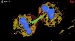
FIGURE 3. 4D image of a dividing cell that was detected by Aivia in a feedback loop with LAS X from Leica Microsystems.
Microscopy enabled by artificial intelligence (AI) will improve life sciences research and development across key therapeutic categories. Promising developments include autonomous...
Leica Microsystems
The Mateo TL digital transmitted light inverted microscope is designed for use in cell culture routine.
Leica Microsystems
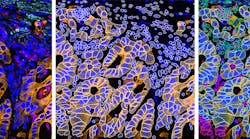
The Cell DIVE scalable multiplexing platform allows researchers to image 60+ biomarkers in a single tissue section.
(Credit: Leica Microsystems/Imperial College London)
Csm Header 32 Imperial Dad5ac29f2
The Leica and Imperial Imaging Hub will provide deeper insights into high-quality images and acquired data through improved optical imaging workflows.
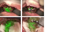
The Leica GLOW800 AR surgical microscope accessory depicts blood flow and related tissue perfusion, which can be useful for the placement and monitoring of an aneurysm clip.
Commercial microscopy developments promise better outcomes for both research and surgical applications.

Leica Microsystems GmbH Categories

Buyer’s Guide Listing Information

Click here for listing information and directions on how to add or update your company.

Request More Information From Leica Microsystems GmbH

By clicking above, I agree to Endeavor Business Media's Terms of Service and consent to receive promotional communications from Endeavor, its affiliates, and partners per its Privacy Notice. I also understand my personal information will be shared with the sponsor of this content, who may contact me about their offerings per their privacy policy. I can unsubscribe anytime.

Additional content from Leica Microsystems GmbH

1912 Lfw Cover Hero
In 2019, the wide-ranging advances in photonics include precision fabrication and measurement, biosensing and imaging, and the use of AI.
This Cell Atlas diagram allows interactive investigation, and it’s backed by a database for exploring details of individual genes and proteins, as well as systematically analyzing transcriptomes and proteomes in broader contexts. (See https://www.proteinatlas.org/about/licence)
Today’s game-changers in optical microscopy are not the usual suspects.
(Photo courtesy of Cleopatra Charalampaki, MD, Ph.D., Professor of Neurosurgery, Department of Neurosurgery, Cologne Medical Center, Germany)
View of an arteriovenous malformation viewed with FL560 fluorescence.
The fluorescence filter provides real-time, high-contrast visualization of both cerebral anatomy in native color and fluorescent blood flow.
(Sample courtesy of Jacco van Rheenen, University of Utrecht, the Netherlands)
Confetti Mouse small intestine expressing CFP, GFP, YFP, and RFP.
The SP8 DIVE tunable deep-imaging system is a spectrally tunable solution for multicolor, multiphoton deep-tissue imaging.
(Credit: Hillman/Li/Targoff, Columbia University)
Beating heart of zebrafish larva (56 hrs. after fertilization). Myocytes are expressing GCaMP (calcium indicator) and dsRed (cell marker). Imaged in vivo using SCAPE at 25 volumes/s, with 335 × 288 × 156 µm field of view.
The agreement will commercialize swept confocally aligned planar excitation (SCAPE) microscopy, which forms 3D images of living samples.
121164 Web
A microscope image injection system overlays virtual reality imaging onto the brain when viewed through the eyepiece during surgery.
Leica Dfc9000 D Mi8 300
The Leica DFC9000 monochrome microscope camera has a highly sensitive third-generation sCMOS sensor.
With super-resolution STED microscopy, details of a cell can be focused down to under 50 nm. This image shows the pores of a cell nucleus (stained green), single protein molecules (red), and fibers of a cytoskeleton (white) in a HeLa cell.
A new core facility focused on bioimaging for cell research will open on February 17, 2016.
Hy Volution 300dpi
The HyVolution true confocal super-resolution microscopy add-on from Leica Microsystems works with the company's TCS SP8 confocal microscope to resolve structures down to 140 ...
The SuperK EXTREME supercontinuum laser powers the Leica TCS SP8 X confocal microscope used for high-resolution bioimaging.
Leica Microsystems and NKT Photonics have signed a supply contract to deliver significant numbers of NKT's supercontinuum lasers.
1509 Lfw Pro 7
The Leica DMC4500 allrounder camera is designed for industrial and life science microscopy.
Leica Em Cryo Clem 300dpi
The Leica EM Cryo CLEM system from Leica Microsystems facilitates correlative light and electron microscopy (CLEM).