Skip to main content
Global coverage of photonics technologies, applications, and innovations
Lasers/Sources
Optics
Quantum
Test/Measurement
Buyers' Guide
Sections
Lasers/Sources
Optics
Quantum
Test/Measurement
Buyers' Guide
Laser Processing
Bio & Life Sciences
Detectors/Imaging
Executive Forum
Special
PhotonicsNXT 2026
2027 Innovators Awards
Issue Archive
Newsletter Archive
Webinars
White Papers
Events
Videos
Podcast
Contact
About Us
Contact Us
Get Published
Advertise
Magazine Subscription
Newsletter Subscription
Affiliated Brands
DATA CENTER FRONTIER
LIGHTWAVE
VISION SYSTEMS DESIGN
ELECTRONIC DESIGN
Follow us on
https://www.facebook.com/pages/Laser-Focus-World/126899915297
https://www.instagram.com/laser.focus.world
https://www.linkedin.com/showcase/laser-focus-world
https://youtube.com/laserfocusworld
Fluorescence
Cameras
LYNRED USA
Offering OEMs and system integrators infrared camera cores, cooled detector assemblies, uncooled thermal sensors. SWaP designs - near IR, SWIR, MWIR, LWIR, VLWIR spectral ranges...
Sept. 1, 2026
(Image credit: Tomocube)
tomocube
Bio&Life Sciences
Bioimaging platform features AI-powered image reconstruction algorithms
March 21, 2025
(Image credit: CRAIC Technologies)
craic_technologies
Detectors & Imaging
Confocal Raman microscope supports multimodal imaging techniques
Dec. 21, 2024
(Image credit: Wei Wei and Mei Xueting)
Depiction of the adaptive sampling scheme, in which a laser beam patterned by a digital micromirror device selectively illuminates neurons and their activity in brain tissue.
Bio&Life Sciences
Two-photon microscope cracks neural networks code
Dec. 13, 2024
(Image credit: Exalos)
exalos
Lasers & Sources
Fabry-Perot laser diodes have use in spectroscopy
Dec. 9, 2024
(Image credit: Leica Microsystems)
leica_microsystems
Bio&Life Sciences
Digital fluorescence microscope offers AI-powered confluency checks
Oct. 30, 2024
(Image credit: Horiba Scientific)
horiba_scientific
Science & Research
Fluorescence lifetime imaging platform adds confocal high-speed laser scanning
Sept. 8, 2024
(Image credit: Prospective Instruments)
FIGURE 1. 3D volume scan of a xenograft (yellow arrow) model in a zebrafish larvae imaged with TPEF and label-free CARS microscopy; the label-free CARS signal is depicted in red and indicated by pink arrow. Scale bar: 100 µm.
Bio&Life Sciences
Bioimaging: Is it possible to improve the performance of microscopes?
Aug. 20, 2024
(Image credit: Excelitas PCO)
excelitas_pco
Detectors & Imaging
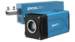
FLIM camera system provides 1008 × 1008 pixel resolution
July 4, 2024
Looking for Something?
Load More Content