Spectroscopy uncovers the hidden in art and archaeology
A wide range of spectroscopies are used in the areas of art and archaeology (A&A), including Raman spectroscopy, laser-induced breakdown spectroscopy (LIBS), x-ray fluorescence (XRF), and reflectance. While most in the laser community don’t think of art conservation, cultural heritage, and archaeological studies as key spectroscopic applications, the history of spectroscopy in these fields dates back more than 40 years.
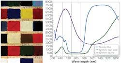
For example, in 1979 the journal Studies in Conservation published a detailed analysis of epoxy resins used in stained-glass conservation, using both UV-visible and infrared (IR) spectroscopy.1 In the same year, an article published in Analytical Chemistry described a technology called the “Laser Raman Molecular Microprobe,” known today as a Raman microscope, listing archaeology as a key application.2 A modern example is shown in Figure 1, where fiber-optic reflectance spectroscopy was used to categorize various pigments.
Today, the worlds of spectroscopy and conservation sciences have become so deeply intertwined that the Society of Archaeological Sciences (SAS) officially became a member of the Federation of Analytical Chemistry and Spectroscopy Societies (FACSS) in 2019. The addition of SAS to FACSS contributed to the decision to select cultural and archaeological analysis as the primary theme of the 2020 SciX conference, taking place October 11-16, 2020. To better understand the growing role of optical spectroscopy in A&A, we interviewed several of the presenting authors in advance of the conference.
Why use spectroscopy?
When asked about the importance of spectroscopy in the field of A&A, McKenzie Floyd, art curator and chemistry educator, replied, “Optical spectroscopy is particularly relevant in the field of A&A research due to the need for noninvasive, nondestructive methods of analysis.” When asked the same question, Moupi Mukhopadhyay, a Ph.D. candidate in the Department of Conservation of Archaeological and Ethnographic Materials at the University of California Los Angeles (UCLA), said, “Practices in archaeology and technical study of art are tied to conservation ethics, which increasingly move towards the principle of minimal intervention.” She went on to add that, “Optical spectroscopy offers options for related studies that can be used to obtain information without engaging in destructive and invasive methods of scientific investigation.”
Each of the presenters interviewed cited nondestructive testing as their primary motivator for using optical spectroscopy. However, the differentiation between destructive and nondestructive testing can have vastly different interpretations within the field. Peter Vandenabeele, professor at Ghent University (Belgium), and Mary Kate Donais, professor at Saint Anselm College (Manchester, NH) and 2020 SciX program chair, discussed this issue extensively in a 2015 review.3
In an attempt to clarify this issue, they defined techniques that cause small damage as “microdestructive.” In comparison, they define nondestructive techniques as “methods that do not consume the sample during the analysis.” For example, IR reflectance spectroscopy would generally be considered a nondestructive technique, whereas LIBS, which relies on laser ablation, would be microdestructive. Raman spectroscopy can fall into either category, depending on the excitation laser wavelength and intensity.
It’s important to point out that, while art and archaeology are often lumped together as a single field, there are differences between the two that affect their tolerance for damage, which in turn dictates the viability of a particular spectroscopic technique. In an interview with Karen Trentelman, Senior Scientist at the Getty Conservation Institute (Los Angeles, CA), she explained that archaeologists are often primarily looking to collect data from a sample to answer a sociological question. Once placed in a museum, the object itself is what is of most importance, not the information in which it contains, therefore “preserving originality” becomes the primary motivation for how they work. The extremely low damage tolerance in art is why microdestructive techniques like LIBS are rarely used in art, but used heavily in archaeology.
Seeing beneath the surface
In addition to analyzing the surface of an object, spectroscopic techniques allow researchers to look under the surface. One of the most stunning examples is Rembrandt’s “An Old Man in Military Costume” (see Fig. 2a). The team at the Getty Conservation Institute, in collaboration with the University of Antwerp (Belgium) and the Delft University of Technology (Netherlands), initially analyzed the painting using mapping-XRF (MA-XRF) to determine the basic structure of the underimage.4Recently, they teamed up with John Delaney at the National Gallery of Art (Washington, DC) to collect hyperspectral IR reflectance images, using a modified near-infrared (near-IR) imaging spectrometer from Surface Optics (San Diego, CA) with a spectral range from 900 to 1700 nm (see Fig. 2b).5 By combining the MA-XRF and hyperspectral image, they improved the contrast of the underdrawing and detected additional sets of eyes (not shown in Fig. 2), indicating that Rembrandt made multiple attempts before presumably giving up and starting over.
Spatially offset Raman spectroscopy (SORS) is another promising method for analyzing subsurface composition of artwork. SORS uses a variable spatial offset between the excitation laser and the collection path to measure the Raman scattering below the surface. SORS is already used in biomedical and security applications, but has only recently been applied to conservation science. In 2014, Claudia Conti and her team at Italy’s National Research Council’s Institute of Heritage Science (ISPC-CNR; Rome), in conjunction with her former advisor Giuseppe Zerbi at Polytechnic University of Milan (Italy) and Pavel Matousek, the inventor of SORS, at Rutherford Appleton Laboratory (Didcot, England), demonstrated a variant of SORS they called micro-SORS for analyzing thin paint layers.6 The following year, the team at ISPC-CNR, again collaborating with Matousek, demonstrated the use of micro-SORS on painted sculptures and plasters.7 In late 2019, Conti et al. published a review of the latest applications of micro-SORS in art analysis.8 These contributions have helped to earn Conti the 2020 Clara Craver award, presented by the Coblentz Society at SciX for significant contributions in applied analytical vibrational spectroscopy.
Spectroscopy in the field
It is not always possible to bring the object to the lab, particularly for archaeological studies. When asked for comment, Mary Kate Donais, who frequently uses portable XRF, Raman, and LIBS, said that “[portable spectroscopy] has with absolute certainty led us to a better understanding of our site and new directions to our work.” In 2016, Mary Kate Donais and her student Luke Douglass presented fieldwork using a SciAps (Woburn, MA) LIBZ500 portable LIBS spectrometer analyzing pottery from two different archaeological sites during Saint Anselm College Field School in Orvieto, Italy.9 This study showed that the differences in the elemental composition implied that some of the ceramics were locally sourced while others came from a different location.
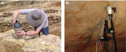
The team from Saint Anselm also used LIBS for analyzing wall mortar at the dig site (see Fig. 3a). Often when performing field analysis, archaeologists may need stability above and beyond that of a handheld device. One example is when Microbeam SA (Madrid, Spain) integrated a portable fiber-optic Raman system from B&W Tek (Newark, DE) coupled to a compact microscope mounted on a motorized tripod to analyze prehistoric cave paintings at the Cueva del Castillo in the region of Cantabria, Spain (see Fig. 3b).Furthering the trend towards portability, in 2019 Alexa Torres and McKenzie Floyd attached a 750 nm cut-on filter to the camera of a Samsung HTC smartphone, creating an extremely low-cost portable near-IR imager.10 With this simple near-IR smartphone device, they were also able to see under images beneath the surface of paint in a way similar to the team at the Getty Conservatory. It is important to note that silicon-based CCD cameras are only capable of detecting wavelengths up to 1100 nm, and they do not capture full spectral data. Still, they were able to detect underimages in multiple portraits, indicating changes the artists made during production.
They also used the near-IR smartphone device to see faded pigments in the mural on an exterior wall. As shown in Figure 4, the blue paint in the center of the mural was significantly faded (see Fig. 4a), but since the blue pigment was so much more absorbent in the near-IR, it is visible in the near-IR filtered image (see Fig. 4b).When asked about the future of optical spectroscopy in A&A, McKenzie Floyd said, “Portability, efficiency, and ease of use are three important values in this field.” She went on to explain that “increasingly small and accessible tools will be deployed to historic and archaeological sites.” When asked her thoughts, Karen Trentelman was excited about the idea of being about to take near-IR images with a smartphone while visiting a gallery. She explained that tools like this would be excellent for collecting the initial data needed to write a proposal to conduct a more detailed secondary analysis. It seems clear from these trends that the future of spectroscopy in A&A will require nondestructive, portable, multimodal instrumentation.
ACKNOWLEDGMENTS
The author would like to thank all of the researchers presenting at the SciX who kindly answered questions both by email and over video chat. He would also like to thank Mary Kate Donais and Rob Lascola, who helped to facilitate these interviews.
REFERENCES
1. N. H. Tennent, Stud. Conserv., 24, 4, 153–164 (1979).
2. P. Dhamelincourt et al., Anal. Chem., 51, 3, 414A–421A (1979).
3. P. Vandenabeele and M. K. Donais, Appl. Spectrosc., 70, 1, 27–41 (2016).
4. K. Trentelman et al., Appl. Phys. A, 121, 3, 801–811 (2015).
5. D. MacLennan et al., J. Am. Inst. Conserv., 58, 1–2, 54–68 (2019).
6. C. Conti et al., Appl. Spectrosc., 68, 6, 686–691 (2014).
7. C. Conti et al., J. Raman Spectrosc., 46, 5, 476–482 (2015).
8. C. Conti et al., J. Cult. Herit. (2019).
9. L. Douglass and M. K. Donais, “Elemental analysis of bucchero pottery using laser induced breakdown spectroscopy,” SciX, Minneapolis, MN (Sep. 18-23, 2016).
10. A. Torres and M. A. Floyd, J. Chem. Educ., 96, 6, 1129–1135 (2019).
About the Author
Robert V. Chimenti
Director, RVC Photonics LLC
Robert V. Chimenti is the Director of RVC Photonics LLC (Pitman, NJ), as well as a Visiting Assistant Professor in the Department of Physics and Astronomy at Rowan University (Glassboro, NJ). He has earned undergraduate degrees in physics, photonics, and business administration, as well as an M.S. in Electro-Optics from the University of Dayton. Over a nearly 20-year career in optics and photonics, he has primarily focused on the development of new laser and spectroscopy applications, with a heavy emphasis on vibrational spectroscopy. He is also very heavily involved in the Federation of Analytical Chemistry and Spectroscopy Societies (FACSS), where he has served for several years as the Workshops Chair for the annual SciX conference and will be taking over as General Chair for the 2021 SciX conference.

![FIGURE 2. Rembrandt Harmensz van Rijn, An Old Man in Military Costume, 1630–31, oil on panel, 65.7 × 51.8 cm, J. Paul Getty Museum 78.PB.246 (a); hyperspectral infrared (IR) image rotated 180° to emphasize the underdrawing (b) [5]. FIGURE 2. Rembrandt Harmensz van Rijn, An Old Man in Military Costume, 1630–31, oil on panel, 65.7 × 51.8 cm, J. Paul Getty Museum 78.PB.246 (a); hyperspectral infrared (IR) image rotated 180° to emphasize the underdrawing (b) [5].](https://img.laserfocusworld.com/files/base/ebm/lfw/image/2020/09/2010LFW_chi_2.5f747c9e32f64.png?auto=format,compress&fit=max&q=45?w=250&width=250)

![FIGURE 4. Unfiltered smartphone image (a) and near-IR smartphone image (b) of a mural on the south exterior wall of Bldg. 4408, Fort Ord [10]. FIGURE 4. Unfiltered smartphone image (a) and near-IR smartphone image (b) of a mural on the south exterior wall of Bldg. 4408, Fort Ord [10].](https://img.laserfocusworld.com/files/base/ebm/lfw/image/2020/09/2010LFW_chi_4.5f747d185e3b3.png?auto=format,compress&fit=max&q=45?w=250&width=250)
