Non-invasive RS-OCT instrument screens for skin cancer
Belfast, Northern Ireland--An Andor near-infrared-enhanced Newton camera was chosen as the core element of a Raman diagnosis module that is part of a portable instrument that combines a Raman spectroscopy (RS) system with an optical coherence tomography (OCT) device to enable sequential acquisition of co-registered OCT and RS data sets. The RS-OCT probe was developed by a joint U.S.-Dutch team under the leadership of Anita Mahadevan-Jansen of Vanderbilt University (Nashville, TN) specifically to perform non-invasive morphological and biochemical characterization of skin cancers.
The most common form of human cancer, skin cancer rates continue to climb from their current estimate of more than three million new cases each year. Although skin cancer can be successfully treated with an early diagnosis and complete resection, the non-optical processes today are invasive, subjective, lengthy, and expensive--requiring expert visual inspection, biopsy, and histopathology.
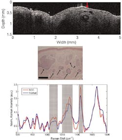
The RS-OCT probe can screen large areas of skin up to 15 mm wide to a depth of 2.4 mm with OCT to visualize microstructural irregularities and perform an initial morphological analysis of lesions. The OCT images are then used to identify locations to acquire biochemically specific Raman spectra.
"Due to the inherently weak nature of Raman scattering and the relatively intense background autofluorescence from tissue, collection efficiency is a critical factor in the design of clinical RS probes," says Chetan A. Patil of Vanderbilt University. "Unlike confocal approaches that emphasize axial resolution, our probe is designed to prioritize collection efficiency. The high sensitivity of the Andor Newton CCD’s back-illuminated, deep-depletion, thermo-electrically cooled design is well suited to the stringent demands of in vivo Raman spectroscopy. The Labview software development kit, which is supplied with the camera and supported directly by Andor, was crucial in the development of our first probe and in our on-going research," adds Patil.
SOURCE: Andor; www.netdyalog.com/news/at/pr11/001.asp?649973-056768-826731-292158
IMAGE: An OCT image (upper) shows a set of hypo-reflective regions in the dermis beneath the red arrow that probably correspond to cancerous tumor cell nests based on the pathology. The tumor cells are also shown in a representative histology (middle; 10x magnification, scale bar = 150 μm). The mean Raman spectra exhibit distinct differences between the cancerous and normal cells in the regions centered at 1090, 1300, and 1440 cm-1. (Courtesy Andor Technology)
