Laser approach promises high-performance, noncontact ultrasound
A non-ionizing technique for fast imaging of soft tissue, ultrasound imaging is currently the most frequently used diagnostic-imaging modality. But it requires physical contact between the probe and the patient’s skin, which introduces unwanted image variability. Now, researchers at the Massachusetts Institute of Technology (MIT; Cambridge, MA) have introduced a laser ultrasound (LUS) technique that promises the safety, accuracy, and convenience of traditional ultrasound without physical contact with the patient—and greater performance, too, including reduced image variability and larger volumetric images without an oversized transducer array.
Sound decisions
An ultrasound image is produced by sending high-frequency sound waves into tissue, then measuring the reflections from various tissue interfaces. A conventional handheld probe contains many piezoelectric transducers that emit and detect high-frequency vibrations. Sonographers apply coupling gel to the skin to ensure proper sound transduction, and generally apply pressure on the skin with the handheld probe. The errors introduced by variations in probe orientation and applied pressure create variability that makes it difficult to perform longitudinal studies or quantitative imaging. And studies indicate that as many as 90% of practicing sonographers suffer work-related musculoskeletal injury from the required application of pressure.
Photoacoustic imaging attempts to bypass those limitations by generating sound waves within the patient’s tissue. The idea is to convert optical energy from a time-varying laser into acoustic energy inside the tissue. When a pulse of light is absorbed by tissue, the tissue heats and expands, creating a pressure wave. The laser can be matched to tissue characteristics in such a way as to produce an ultrasound signal originating from the tissue itself. Ultrasonic detectors on the skin surface detect the transmitted waves, which are then reconstructed into a tissue image. But incident light is not absorbed equally within the tissue. To some degree, the optical wavelength can be selected to target different types of tissue at different depths, but the relevant wavelengths of light used are strongly attenuated in biological tissue.
The MIT team developed an ultrasound-generation technique that combines the advantages of both traditional ultrasound and photoacoustic imaging. They chose a 1540 nm pulsed laser as the excitation source, a wavelength that is absorbed strongly by biological tissue. That means the photoacoustic source is located just within the surface of the skin, localized similarly to traditional ultrasound. In contrast to traditional ultrasound, however, the laser source requires no skin contact, similar to photoacoustic imaging.
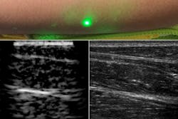
The researchers replaced the acoustic detector of conventional photoacoustic imaging with a 1550 nm continuous-wave laser-Doppler vibrometer to measure skin vibrations (see figure). This makes for a fully noncontact system, nearly eliminating the risk of repetitive-stress injuries for sonographers. The selection of wavelengths around 1550 nm allows the system to take advantage of advances in telecommunications technology, as well as matching tissue characteristics. In addition, the power and energy required by the LUS system are well within laser-exposure safety limits for human use.
Early-stage results
The researchers validated their method on tissue phantoms with embedded metal features, porcine tissue, and human forearms, where the reconstructed images were consistent with traditional ultrasound images. The LUS images are comparable to circa-1960s ultrasound, prior to the electronics and manufacturing improvements that led to the rapid, high-quality imaging of contemporary ultrasound. Xiang (Shawn) Zhang, a researcher and author on the MIT team, noted that “early-stage ultrasound images were generated using a single transducer that was manually moved to generate an image, which is very similar to our present LUS, since we are moving laser spots on the skin surface. The system should benefit from an innovation pathway similar to that responsible for the past 50 years of traditional ultrasound development.” Advances in silicon photonics, for example, could lead to the development of multipoint light sources comparable to the piezoelectric arrays of traditional ultrasound.
LUS is poised not just to match traditional ultrasound, however, but to exceed its capabilities. Since light can be steered anywhere on the skin surface, LUS imaging is not constrained by the physical dimensions of a traditional ultrasound probe. This makes it possible to create larger volumetric images without a prohibitively large piezoelectric array. With LUS, the propagation of the sound also doesn’t depend on the orientation of the light and no contact pressure is applied, so overall image variability can be reduced.
For now, Zhang hopes the first LUS human images will lead to “broader interest in the LUS technique and bring together experts in related fields to overcome the challenges as the technology develops toward clinical translation.”
About the Author
Richard Gaughan
Contributing Writer, BioOptics World
Richard Gaughan is the Owner of Mountain Optical Systems and a contributing writer for BioOptics World.

