Ultrafast two-photon microscope to delve into neurological diseases
A team of researchers at The Chinese University of Hong Kong (CUHK) has developed a digital holography-based (DH) two-photon excitation (TPE) microscope to generate simultaneous video-rate fluorescence imaging and multipoint optical stimulation. This allows the tracking of nerve cells activities and thus may help the study of neurological diseases, such as glaucoma.
Related: Raman imaging method could better understand neurodegenerative diseases at the molecular scale
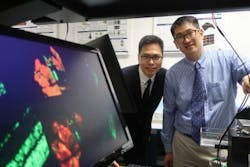
By scanning the retinal ganglion cells using the DH-TPE microscope, scientists can understand the molecular mechanisms of optic nerve degeneration in glaucoma and find ways to tackle it. Research team leader Shih-Chi Chen, associate professor in the Department of Mechanical and Automation Engineering at CUHK, is now working on this project with Prof. Christopher Kai Shun Leung, a professor in the Department of Ophthalmology and Visual Sciences at the Faculty of Medicine at CUHK, and the Hong Kong Eye Hospital.

Using the DH-TPE microscope, Prof. Shih-Chi Chen from the Faculty of Engineering and Prof. Christopher Kai Shun Leung from the Faculty of Medicine scan retinal ganglion cells of living animals to find out the molecular mechanisms of optic nerve degeneration in glaucoma.
The DH-TPE microscope is powered by a digital micromirror device (DMD), a chip that contains millions of micromirrors switching at tens-of-kilohertz speed. By controlling the amplitude and phase of the input laser via binary holograms and the fast-switching micromirrors, the laser beam can be split into up to 20 focal points for simultaneous optical stimulation and real-time fluorescent imaging. Each focus can be independently controlled to scan along arbitrarily defined paths or surfaces at 22.7 kHz. More importantly, the DMD scanner is also an ultrafast beam shaper, and by superposing the scanning and wavefront-correction holograms, the point spread function can be engineered or even shaped into other novel beam modes to achieve efficient 3D imaging. The DH-TPE microscope's imaging functionalities include random-access imaging, multiplane imaging, a 3D programmable imaging plane, point-specific wavefront correction, and simultaneous video-rate fluorescent imaging and multipoint optical stimulation.
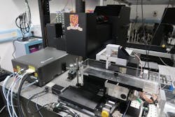
The DH-TPE microscope is powered by a digital micromirror device; it can generate simultaneous video-rate fluorescent images, which allow the tracking of nerve cells' activities.
Through funding support from the Innovation and Technology Commission (ITC), Chen's team is setting up the DH-TPE microscope at the Hong Kong Eye Hospital, collaborating with a team led by Leung. The two teams are working together to exploit the unique capability of the DH-TPE microscope to study and understand the basic mechanisms of a few important diseases.