Evident

48 Woerd Ave
Waltham, MA 02453
United States
800-225-8330

More Info on Evident

At Evident, we are guided by the scientific spirit—innovation and exploration are at the heart of what we do. Committed to making people’s lives healthier, safer and more fulfilling, we support our customers with solutions that solve their challenges and advance their work; whether it’s researching medical breakthroughs, inspecting infrastructure, or exposing hidden toxins in consumer products.

Products

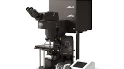
Fv3000
The FLUOVIEW™ FV3000 series of confocal laser scanning microscopes meets some of the most difficult challenges in modern science. Featuring the high sensitivity and speed required...
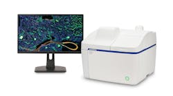
Apx100 With Monitor
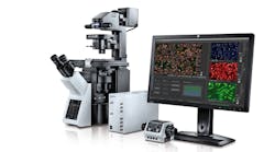
The APEXVIEW™ APX100 all-in-one microscope makes it fast and simple to acquire expert-quality microscope images. Built with renowned Olympus optics, an intuitive user interface...
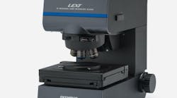
Ols5100
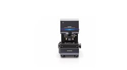
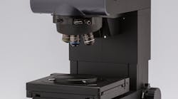
The LEXT™ OLS5100 3D Laser Scanning Microscope is a powerful tool for material analysis. With exceptional measurement accuracy and optical performance, this laser microscope is...
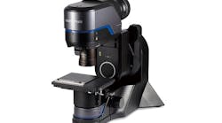
Product Dsx1000 Entry 01
The DSX1000 Digital Microscope is a powerful, fast, and reliable failure analysis tool. With guaranteed accuracy and repeatability, this microscope features a telecentric optical...
Product Szxar1 Overview01
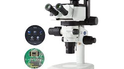
The SZX-AR1 Augmented Reality Microscope System microscope system overlays text and digital images over the field of view, making it easy to follow instructions, read notes, and...

Press Releases

Screen Shot 2023 05 10 At 3 02 04 Pm
Scientific Solutions business splits from global medtech company TOKYO, Japan (April 4, 2022)—Olympus Corporation (“Olympus”) announced the completion of the separation of its...

Articles

(Image credit: Evident)
evident
The DSX2000 fully motorized digital microscope is designed for R&D, quality control, and failure analysis inspection workflows.
(Image credit: Evident Scientific)
evident
The IXplore IX85 automated inverted microscope system provides a 26.5-mm field of view on two integrated imaging ports.
(Sample courtesy of Aoi Gohma and Atsushi Miyawaki, RIKEN CBS-EVIDENT Open Collaboration Center)
FIGURE 1. A 3D image of a live mouse brain acquired using an Evident FV4000 MPE microscope and a 25x TruResolution objective.
Near-infrared (NIR) imaging can help mitigate the challenges of using visible light as an excitation source for biological research.
(Image credit: Evident)
evident
The IPLEX TX II videoscope combines the versatility of a 2.2-mm-diameter flexible scope with an ultrathin 1.8 mm rigid scope in a single package.
evident
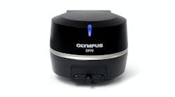
The DP75 digital microscope camera captures images from brightfield to wide-wavelength fluorescence and near-infrared using a single camera.
Evident
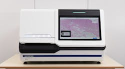
The SLIDEVIEW DX VS-M1 whole-slide imaging system is a digital pathology solution that delivers microscope-quality slide images onscreen.
FIGURE 1. The Olympus eyepoint height adjuster can be adjusted from 30 to 150 mm, allowing users of any height to operate the scope comfortably.
Incorporating proper ergonomics into your industrial microscope setup and routine helps to prevent injuries and increase productivity.
Photo 236540213 © Marek Uliasz | Dreamstime.com
Dreamstime Xl 236540213
Business announcements were plentiful in February 2022 for the optics and photonics industry, spanning several application and product markets.
Olympus
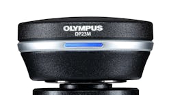
The DP23M monochrome microscope camera features a backside-illuminated monochrome CMOS sensor with 2 × 2 binning.
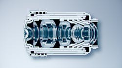
FIGURE 1. Example of a complex and compact group of lenses inside an objective.
New objective lens technology delivers high levels of numerical aperture, chromatic aberration correction, and flatness at the same time for improved optical performance.

Buyer’s Guide Listing Information

Click here for listing information and directions on how to add or update your company.

Request More Information From Evident

By clicking above, I agree to Endeavor Business Media's Terms of Service and consent to receive promotional communications from Endeavor, its affiliates, and partners per its Privacy Notice. I also understand my personal information will be shared with the sponsor of this content, who may contact me about their offerings per their privacy policy. I can unsubscribe anytime.

Additional content from Evident

Photo 88499141 © Korn Vitthayanukarun | Dreamstime.com
dreamstime_xxl_88499141
Here are the top four photonics business moves that made headlines during the week ending December 1, 2023.
(Photo: ID 60933364 © Pichetw | Dreamstime.com)
Dreamstime M 60933364
Quest Photonic Devices’ multispectral imaging offerings will drive the growth of Olympus’ surgical endoscopy business.
FIGURE 1. The effect of laser wavelength on lateral resolution: A grating with 260-nm-wide lines imaged using a laser scanning confocal microscope with a 658 nm laser (a) and a grating with 120-nm-wide lines imaged using the Olympus LEXT OLS5000 laser scanning confocal microscope with a 405 nm laser (b) are shown.
For industrial surface measurement, a laser scanning confocal microscope can provide high accuracy and the right roughness parameters for your application.
Olympus
The LEXT OLS5100 3D laser scanning microscope is designed for failure analysis and material engineering research with 0.12 µm lateral resolution.
Olympus Cropped
The scanR high-content screen (HCS) station version 3.2 incorporates Olympus X Line objectives for improved chromatic aberration correction, image flatness, and resolution.
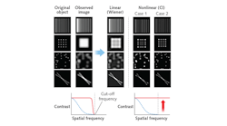
FIGURE 1. Results from linear and nonlinear deconvolution are shown in these images depicting original objects before image acquisition, observed confocal image through microscope, and following deconvolution (left). The center panel depicts linear deconvolution with a Wiener filter, resulting in ringing artifacts observable in the beads and crossed filaments. Nonlinear deconvolution results (right) improve image quality, but may create data beyond the cut-off spatial frequency. Case 1 results show artifacting in connections between adjacent beads. When deconvolution parameters are carefully adjusted (as depicted in Case 2 column), good results can be achieved.
A combination of advanced algorithms and improved hardware is driving the popularity of deconvolution for optical confocal microscopy.
(Image credit: Dan Houchin, Olympus America)
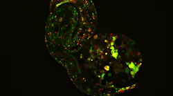
FIGURE 1. Superresolution techniques, enabled by software and hardware advances, highlight the most minute details and processes that define life; an example is this image depicting early embryonic development captured on the Olympus SpinSR superresolution microscope.
As technologies have progressed, certain types of superresolution optical imaging—so-called functional superresolution techniques—have become more user-friendly across a broad...
1712 Lfw Pro 12
The LEXT OLS5000 3D laser confocal scanning microscope includes 4K scanning technology.
Courtesy of Olympus
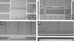
These images were acquired using various Olympus IR objective lenses, a BX2M-IR upright compound microscope, an XM10-IR digital camera, and Olympus Stream software: 10x (a), 20x (b), 50x (c), and 100x (d).
Imaging through silicon with dedicated IR objective lenses enables nondestructive flip-chip analysis and other essential evaluations.
Olympus
Content Dam Lfw Online Articles 2017 11 Olympus
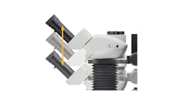
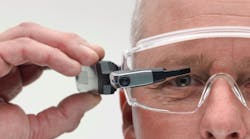
The device uses "pupil division" to imitate a semitransparent viewing screen.
(Courtesy of BaySpec)
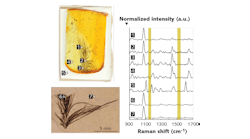
FIGURE 1. Confocal measurements of fossil feathers embedded in epoxy and amber show keratin, but no carotenoid (yellow) contributions in their Raman spectra.1 The measurements were done with a BaySpec Nomadic confocal microscope (bottom) at 1064 nm, one of the microscope's three Raman excitation wavelengths (the other two are 532 and 785 nm).
When paired with a high-resolution microscope, Raman spectroscopy opens a world of knowledge for scientists, doctors, and industrial researchers.
1601 Lfw Pro Add 1
The DSX500 digital microscope observes, measures, and analyzes electronic components, circuit boards, construction materials, semiconductors, medical devices, and shielding components...