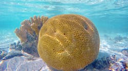
University of Copenhagen (Denmark), University of Technology Sydney (Australia), and Oregon Health University (USA) scientists are using a well-known biomedical imaging technique called optical coherence tomography (OCT) to obtain breakthrough insights to the structural organization and dynamics of reef-building corals. Their results have been published in the multidisciplinary Journal of the Royal Society Interface.
RELATED ARTICLE: Optical Coherence Tomography: Advances in functional OCT
Direct observation of living corals is not easy and has relied on bright field imaging and epifluorescence microscopy with limited depths and resolution due to the opaque coral tissue, which is composed of different cell layers and exhibits diffuse backscatter from the underlying coral skeleton. The use of visible light for such observations can also negatively influence the corals by stimulating photosynthesis or exposing them to potentially harmful UV and blue light.
An international team of scientists headed by professor Michael Kühl at the Department of Biology, University of Copenhagen has now surpassed such limitations in observing the tissue organization of living corals by using OCT.
Kühl explains, "OCT is an optical ultrasound-like technology that is e.g. employed by doctors to monitor tissue damage in the eye. It involves the use of non-actinic near-infrared radiation that penetrates deeper into tissue than visible light and can reveal microscopic structures with different reflective properties. We used an OCT system that enabled rapid 3D scanning of a 1-2 cm2 area down to a tissue/skeleton depth of 1-3 mm at a spatial resolution of a few µm. This enabled fascinating insights to the internal and external tissue-organization over the skeleton of living corals." See video:
It was possible to identify different tissue layers and quantify their plasticity upon changes in light exposure on living corals. Corals rapidly contracted their tissue under high light stress, making it more reflective thereby protecting their symbionts against excess light. OCT also enabled the quantification of fluorescent host pigments organized in granules that also made the tissue more reflective especially after contraction.
In the dark, corals expand their tissues to gain better access to oxygen, and OCT showed that the tissue surface area of corals can be doubled at nighttime. The surface area of corals exposed to seawater and incident light is thus very dynamic, and OCT can now quantify such changes. This can have important implications for the measurements of coral metabolic rates, which typically are normalized to the surface area of the coral skeleton after the tissue has been removed - assuming that such area measurements are representative of the coral tissue surface area. The OCT results indicate that this assumption needs revision.
It was also possible to monitor the production of coral mucus on the tissue surface, which is an important component of coral life as mucus harbors beneficial microorganisms and also traps particles for feeding or self-cleaning purposes. Enhanced mucus production is also a signature of stressed corals, e.g. upon onset of coral bleaching. Furthermore, corals can expand special defensive tissue structures such as mesenterial filaments upon mechanical stress, and OCT could also visualize such dynamic responses.
SOURCE: University of Copenhagen; http://news.ku.dk/all_news/2017/03/breakthrough-in-live-coral-imaging/
About the Author

Gail Overton
Senior Editor (2004-2020)
Gail has more than 30 years of engineering, marketing, product management, and editorial experience in the photonics and optical communications industry. Before joining the staff at Laser Focus World in 2004, she held many product management and product marketing roles in the fiber-optics industry, most notably at Hughes (El Segundo, CA), GTE Labs (Waltham, MA), Corning (Corning, NY), Photon Kinetics (Beaverton, OR), and Newport Corporation (Irvine, CA). During her marketing career, Gail published articles in WDM Solutions and Sensors magazine and traveled internationally to conduct product and sales training. Gail received her BS degree in physics, with an emphasis in optics, from San Diego State University in San Diego, CA in May 1986.
