Existing diagnostic tools that examine and image the retina are suitable enough for use with adults, but not for infants and toddlers. Current tools of this sort put demands on both the patient (who must sit still and focus for periods of time) and the clinician (even handheld versions weigh several pounds, making them hard to use). Also, none provide resolution sufficient to see individual photoreceptors.
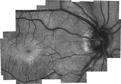
An interdisciplinary research team at Duke University (Durham, NC) has developed a lightweight, miniaturized device able to capture retinal images with cellular resolution.1 About the size of a cigarette pack and weighing just over three ounces, it combines scanning laser ophthalmoscopy (SLO) and optical coherence tomography (OCT) in the first device ever to directly measure the density of "cone" photoreceptors (which convert light into nerve signals) in infants (see figure).
As such, the instrument promises to enable assessment of the impact of injury, as well as "new research that will be key in future diagnosis and care of hereditary diseases," according to engineering professor and OCT pioneer Joseph Izatt, who co-led the collaborative research group along with ophthalmology and biomedical engineering professors Sina Farsiu and Cynthia Toth.
Optical design
Keys to the design, says first author Francesco LaRocca, are 1) a single light source for both SLO and OCT; 2) a compact scanning mirror (Mirrorcle Technologies); 3) custom optics and mechanics (which he and fellow Duke PhD Derek Nankivil designed); and 4) a novel compact telescope design that relays light from the scanning mirror to the eye's pupil using converging rather than collimated light at the input.
The probe incorporates an off-axis parabolic mirror that collimates light from an optical fiber, while an achromatic lens focuses the light before the scanning mirror. Commercially available and custom-designed lenses work together to magnify the beam: A plano-concave lens adjacent to the probe's intermediate focal plane substantially removes field curvature. A custom biconvex lens (optimized using Zemax ray-tracing software for curvature, thickness, glass type, and position) keeps the principal plane of the plano-concave and biconvex lens pair a focal-length distance from the scanner. Finally, an asymmetric triplet, designed to provide magnification of 2.8, serves to minimize induced monochromatic aberration, while also correcting chromatic aberrations across the light spectrum. An accurate eye model, spanning a 7° field of view (FOV), was critical for designing the custom optics.
Capability and impact
Through testing on adults, the new device proved capable of accurately capturing photoreceptor density information. It was also used for imaging in children who were already having an eye exam under anesthesia. "But because children have never been imaged with these systems before, there's no gold standard that we can compare it to," LaRocca said. Indeed, lacking such a measurement tool, little is known about development of the retina—which is mature by the time a child reaches age 10. LaRocca notes, however, that the team's results "match theories of how cones migrate as the eye matures. The tests also showed different microscopic pathological structures that are not normally possible to see with current lower-resolution, clinical-grade handheld systems."
With the prototype being used by clinicians at Duke Health, the amount of information being gained from children's scans could eventually create a database to give a much better picture of how the retina matures with age. Since publication of the paper, the researchers have developed an updated design based on clinician feedback. The new probe provides motorized focus adjustment, a FOV ~5 times larger than the original, and, because it uses swept-source OCT, is faster (though in this version, OCT is not augmented by SLO because of design tradeoffs between resolution and FOV).
REFERENCE
1. F. LaRocca et al., Nature Photon., 10, 580–584 (2016).

