In optical coherence tomography (OCT) imaging, a high lateral resolution typically means a short depth of field or reduced axial resolution. In fact, the depth of focus is given by twice the Rayleigh range, where the Rayleigh range is proportional to the square root of the lateral resolution. In order to image at the cellular level (several microns), the depth of field is reduced to tens of microns, limiting the ability of OCT to perform diagnostic functions for thicker biological tissues.
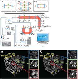
In April 2013, researchers at the VU University Amsterdam (Amsterdam, Netherlands) modified a standard OCT system through an adaptive-optics method called depth-encoded synthetic-aperture OCT, in which pupil segmentation by an annular phase plate (APP) segments the pupil into two parts, creating three distinct optical apertures that can extend the depth of field by a factor of five for focused analysis of 0.3-mm-thick tissues (see figure).1
Basically, the three apertures are encoded to different depths in a single OCT scan, allowing the phase and complex amplitude of three images to be digitally refocused into a single image with improved focus or extended depth of field. However, this earlier research used a laser with a relatively short depth range of only 5 mm; more recently, the use of a laser with a much longer depth range (12 mm) further refines the technique, with depth of focus extended to around 1.2 mm—allowing analysis of tissues four times thicker than the prior result.2
Synthetic-aperture setup
Using a 1310-nm-emitting vertical-cavity surface-emitting laser (VCSEL) with a 75 nm 3 dB bandwidth, 12 mm coherence length, and 200 kHz sweep rate, the beam is split in two with 10% of the beam sent to a fiber Bragg grating (FBG) to create a trigger signal and the other 90% input to the interferometer portion of the setup.
In the interferometer sample arm, the light is collimated to a 3.4-mm-diameter beam, and an objective lens reduces it to a 10.1 μm focal spot and a depth of field of roughly 83.7 μm. The back focal plane of the objective is then sent to a galvanometer mirror and then into an 8-mm-thick polycarbonate (refractive index of 1.56) phase plate with a 1.8-mm-diameter central aperture. The galvo mirror is driven with a sawtooth waveform for raster scanning, allowing linear sampling of the spectrum and direct Fourier transform.
In the digital-refocusing algorithm, the Fourier transform of the OCT signals is computed for the three separate aperture images, creating three complex images representing the backscattered electrical fields. These three images can be coherently added to generate a single image with an optimized focus over an extended depth of field.
Experimental analysis of four slices of melamine spheres fixed in a silicon elastomer curing agent with a total thickness of 1.2 mm proves that the synthetic aperture method can adequately focus spheres throughout the entire thickness as opposed to unfocused results from a standard OCT scan.
“The depth-encoded synthetic-aperture method is ideally suited for implementation in catheters that enter the human body,” says Johannes F. de Boer, director of the LaserLaB and professor in the Physics department at VU University. “A small annular phase plate is easy to integrate into the optical fibers of small catheters. Refocusing the image at the catheter tip does not require any moving parts because the signals of the different apertures are encoded at different depths in the OCT signal. This enables better lateral resolution imaging over large depths in endoscopic OCT.”
REFERENCES
1. J. Mo et al., Opt. Express 21, 8, 10048 (2013).
2. J. Mo et al., Opt. Express 23, 4, 4935 (2015).

