Spectroscopy/Oncology/Optical Biopsy: Visible resonance Raman improves tumor identification and grading

Optical spectroscopy, including Raman methods, offers many advantages over standard techniques for tissue assessment, including noninvasive, label-free, and real-time operation; time efficiency; and reproducibility. Similarly, resonance Raman (RR) spectroscopy shows advantages over conventional Raman using slight absorption from specific biomolecules in the cells and organelles—such as from proteins, lipids, flavin adenine dinucleotide (FAD), amino acids, reduced nicotinamide adenine dinucleotide (NADH), collagens, elastin, carotenoids, hemeproteins, and mitochondrial cytochromes—these molecules contain fluorophores whose absorption of light leads to resonance enhancement of specific vibrational modes (for example, stretching, bending, and scissoring). The spectra collected from resonance-enhanced molecules can be detected at concentrations <1.0 nM. RR can preferentially target the activities of particular molecular species, making Raman peaks more accessible for identifying changes correlated with various human diseases.
A 2019 book, Neurophotonics and Biomedical Spectroscopy, features exploration of RR for detecting human brain tumor (see Fig. 1),1 and new results obtained by the authors, along with colleagues in China and the U.S., are the first to use visible resonance Raman (VRR) spectroscopy to target glioma, which is the most common type of central nervous system (CNS) tumor.2 Since 2011, researchers from the U.S. and China, respectively, have been collaborating on using VRR to study a variety of human tissues. The work is led by the Alfano group at The City College of New York of the City University of New York, which has done pioneering work since 1987 in applying Raman for optical biopsy. The research work was completed by Zhou’s neurological research team in the Beijing laboratory of Tianjin Raman Medical Technology Center (TRMTC) in China.3 There is collaboration between China and the U.S. to solve the cancer problem of the breast and brain.
Glioma and spectroscopy
Even using a combination of state-of-the-art treatment modalities, mortality and recurrence rates are rising for glioma. Although multiple factors affect outcomes for glioma patients, the ability to accurately delineate tumor boundary for effective resection is critical. So is the grading of gliomas, an active area of research that is expected to improve diagnosis and treatment.
The current gold standard for brain tumor diagnosis—biopsy and histopathology—is an invasive and time-consuming process. Alternative detection methods exist, but each has drawbacks. A review of postoperative MRI results revealed that the accuracy and timeliness limitations restrict full glioma resection to only ~65%. Other studies show that in 75% of cases, there was no ability to clearly distinguish among higher- and lower-grade gliomas.
The high cost of emerging molecular genetic pathology, and the desire to increase accuracy, while reducing time-to-diagnosis, are motivating researchers to explore optical molecular histopathology techniques.
Early studies of RR used ultraviolet (UV) light, usually at <300 nm. VRR, on the other hand, uses a 532 nm laser to enhance the Raman signal intensity from intrinsic molecules by ~100X due to its electronic resonance effect inside the tissue. VRR’s signal-to-noise ratio (SNR) is higher, which makes it possible to directly compare cancerous and normal tissues. There is a large background “wing” that needs to be removed. At the same time, VRR uses less laser power and a shorter integration time to collect signals. Therefore, it is safer for application on live tissues and cells.
Grading and margin assessment
The new article reported the application of VRR to evaluate biomarkers for detecting glioma margins and to investigate correlations between biomarkers and tumor grades. After collecting spectra from various grades of glioma tissues, the team applied machine-learning techniques to classify the specimens, and compared the results to results obtained from traditional histopathology. Analyses based on the measured VRR spectral data revealed that the major molecular biomarkers (including carotenoids, tryptophan, amides, proteins, and lipids) are involved in metabolic processes in human brain, and can form the foundation of an optical pathology method.
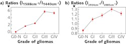
Traditionally, gliomas are classified into grades I to IV according to morphology, degree of malignancy, and location of tumor cells. The study revealed that all RR molecular fingerprints occurring in grade I glioma tissue showed a distinct shift in higher grades of glioma tissues (see Fig. 2).
The team established criteria for differentiating tissue types and used these criteria to identify tumor margins, reporting sensitivity at 100% and specificity of 71% compared to chemical reagent-based histopathological analysis.
Next steps
The team continues to make progress in developing and applying the Raman scattering microscopy techniques in studying brain cancer. Specifically, plans are underway to refine the approach by exploring issues observed in the research (for example, 1588 cm-1 Raman mode) and by identifying real-time correlations between laboratory-derived Raman spectral characteristics and results obtained through examinations at the Beijing laboratory of TRMTC.
The team is also working on multiple instrumentation designs, including a fiber-optic VRR system that measures all molecular spectral biomarkers in real time to detect brain cancer and identify the tumor boundaries in vivo. Moreover, they plan to develop a nonlinear optical microscopy system that will combine VRR and stimulated Raman scattering (SRS) for general optical biopsy applications.
![FIGURE 3. This pre-commercial VRR portable analyzer (model LRR2000 by Jiangsu Raman Medical Equipment Co., Ltd. [JRME Co.] in China), with its fiber-optic probe delivering 532 nm light, produced an accurate diagnosis of brain glioblastoma multiforme (GBM) when used in vivo in a mouse model. The screen provides intraoperative guidance by displaying signals, test results, flickering colors, and ringtones in real time.](https://img.laserfocusworld.com/files/base/ebm/lfw/image/2019/10/1910LFW_alf_3.5d9ce4f57c16f.png?auto=format,compress&fit=max&q=45?w=250&width=250)
A portable VRR device with a scanner for imaging and with integration time <1 s (see Fig. 3) has already been tested on mice and is currently being used to measure human samples ex vivo. In the animal-model experiments reported at the Frontiers in Optics + Laser Science conference (held Sep. 15-19, 2019, in Washington, DC; paper JW3A.5), the laser power was maintained at 3.5 mW. The final spectral resolution was 8 cm-1 in the range of interest (200–4000 cm-1). The 532 nm excitation beam directly excites the surface of the mouse brain (can be tumor or normal brain tissue) for 1 s to obtain a Raman spectrum. A fiber-optic probe with a 200 µm focus spot was used to focus the laser beam and collect the scattered signals from the sample surface—in this case, in vivo mouse. The next step is to apply it on human brain in vivo.
REFERENCES
1. Y. Zhou et al.,“Visible resonance Raman spectroscopy in human brain tissues,” Neurophotonics and Biomedical Spectroscopy, R. R. Alfano and L. Shi (Eds.), 65–106, ISBN 9780323480673. Elsevier, Cambridge, MA (2019); https://doi.org/10.1016/B978-0-323-48067-3.00004-4.
2. Y. Zhou et al., J. Biomed. Opt., 24, 9, 095001 (2019); doi:10.1117/1.jbo.24.9.095001.
3. Y. Zhou et al., J. Biomed. Opt., 17, 11, 116021 (2012).
About the Author
Yan Zhou
Yan Zhou is in the Department of Neurosurgery at the Air Force Medical Center of PLA (Beijing, China).
Shengjia Zhang
Shengjia Zhang is at JRME Co. in China.
Binlin Wu
Binlin Wu is an Assistant Professor in the Physics Department and CSCU Center for Nanotechnology at Southern Connecticut State University (New Haven, CT).
Cheng-Hui Liu
Cheng-Hui Liu is a cancer researcher at the Institute for Ultrafast Spectroscopy and Lasers at the City University of New York (CUNY; New York, NY).
Robert Alfano
Robert Alfano is a distinguished professor of science and engineering at the City College of the City University of New York (CUNY), where he is the director of Institute for Ultrafast Spectroscopy and Lasers. He works primarily in the field of biomedical imaging and spectroscopy, and is known for discovering the white light supercontinuum laser. He has published more than 700 papers in referred journals and has over 100 patents, and has won numerous awards including the SPIE’s inaugural Britton Chance Biomedical Optics Award.
Lingyan Shi
Dr. Lingyan Shi is currently an Associate Professor in the Shu Chien-Gene Lay Department of Bioengineering at UC San Diego. Her research focuses on developing high resolution optical spectroscopy and imaging platforms, and its applications for studying metabolic dynamics in aging and diseases. She discovered the “Golden Window” for deep tissue imaging and developed bioorthogonal metabolic imaging platforms that combine deuterium probing and stimulated Raman scattering (DO-SRS using heavy water and STRIDE with D-glucose) for visualizing metabolic activities in situ. The Shi group transformed SRS into a super resolution microscopy with chemical selectivity by developing Adam optimization-based Pointillism Deconvolution (A-PoD) methods. Dr. Shi holds six awarded patents. She won the Blavatnik Regional Award for Young Scientist in 2018; the Hellman Fellowship Award 2021; the “Rising Star Award” by Laser Focus World, and the “Rising Star Award” by Nature Light Science & Applications in 2021; the “Advancing Bioimaging Scialog Fellow” by RCSA and the Chan Zuckerberg Initiative in 2021, 2022, and 2023; and the Sloan Research Fellow Award in Chemistry 2023.
Dr. Shi has been mentoring graduate and undergraduate students to help them achieve excellence in academic work and become successful engineers and scientists. She plans to continue making additional contributions by enhancing more participation of underrepresented groups from the UC San Diego communities. She has been teaching core undergraduate and graduate courses in the Shu Chien-Gene Lay Department of Bioengineering at the UC San Diego Jacobs School of Engineering.