Recent innovations in nonlinear optics and imaging enable highly efficient microscopic analysis. But science has been unable to realize the potential of such methods because truly taking advantage of them requires a mind-bending application of real-time single-exposure imaging at extraordinarily high temporal resolutions. “While the concept of focusing usually applies to the spatial domain, it is equally applicable to the time domain,” writes a team of North American researchers describing a new technique for imaging the focusing of ultrashort light pulses that meets these demands. Their solution has staggering implications for biomedical imaging, as well as for materials science and other applications.
Temporal resolution saves specimens
Capturing phenomena such as the travel of light through a medium is a monumental challenge that some techniques address by making repeated measurements. But with fragile samples, repeated application of laser pulses can prove devastating: Even laser-engraved glass requires picosecond imaging because it can tolerate no more than a single ultrashort pulse.
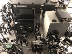
A team led by Lihong Wang, Bren Professor of Medial Engineering and Electrical Engineering at California Institute of Technology (Caltech; Pasadena, CA) and the Director of Caltech Optical Imaging Laboratory (COIL, also in Pasadena), used previous work on single-shot compressed ultrafast photography (CUP), which delivers 100 billion frames per second (fps), as a foundation for its single-shot approach. But to take advantage of the shorter pulses of a femtosecond laser, the researchers developed the T-CUP system (see figure). “We knew that by using only a femtosecond streak camera, the image quality would be limited,” Wang says. “So to improve this, we added another camera that acquires a static image.”
The T-CUP concept is embodied in a system that operates using a type of data acquisition commonly used in applications such as tomography, along with image reconstruction. “Combined with the image acquired by the femtosecond streak camera, we can use what is called a Radon transformation to obtain high-quality images while recording 10 trillion fps,” Wang explains.
To acquire data, a beamsplitter captures the intensity distribution of a 3D scene to create two images. A 2D imaging sensor records the first image directly using spatiotemporal integration. A pseudorandom binary pattern encodes the second image spatially, and a femtosecond shearing unit shears temporal frames onto a single spatial axis. Another 2D imaging sensor records the spatially encoded and temporally sheared frames to form a time-sheared view.
Among key components in the T-CUP system is a CCD camera that performs spatiotemporal integration, and a digital micromirror device (DMD) that accomplishes spatial encoding. Time-varying voltage enables femtosecond shearing when applied to the sweep electrodes of a femtosecond streak camera. And a two-view reconstruction algorithm based on compressed-sensing enables recovery of the dynamic scene being imaged.
The system can image a dynamic scene with 450 × 150 pixels in 350 frames during a single camera exposure. The streak camera’s temporal shearing velocity and the internal CCD’s pixel size, along with temporal shearing direction, together determine the reconstructed video’s frame rate, which can be adjusted from 0.5 to 10 Tfps by varying the shearing velocity. With the combination of its single-shot data capture and its super-fast tunable frame rate and sequence depth (frames per movie), T-CUP can image nonrepeating ultrafast transient phenomena that occur over a wide range of timescales.
Instrumentation implications
In breaking new ground for real-time imaging, T-CUP represents a paradigm shift for analysis of interactions between light and matter. Thus, it portends a new generation of microscopy systems for applications in biomedicine and materials science as well as other applications.
Biomedicine is currently hampered by the fact that living tissue is a dynamic scattering medium with speckle decorrelation on the order of milliseconds. Because existing imaging methods have limited wavefront characterization speeds, deep-tissue spatiotemporal focusing has been realized only in static specimens. But the researchers demonstrated that T-CUP is able to perform femtosecond imaging of transient light patterns in in vivo tissue in single exposures. Integrating T-CUP with interferometry enables examination of a broadband beam’s scattered electric field, which in turn promises to enable spatiotemporal focusing with phase conjugation in living biological tissue. For this reason, the researchers’ work has important implications for biomedical instrumentation: It promises to facilitate widefield two-photon microscopy, as well as advances in photodynamic therapy and optogenetics.
While the T-CUP camera set a record in its initial application by capturing a single femtosecond laser pulse’s temporal focusing in real time—detailing the shape, intensity, and inclination angle of the pulse—the team already sees potential for greater speed. Lead author Jinyang Liang, professor and ultrafast imaging specialist at Canada’s Institute National de la Recherche Scientifique (INRS, Quebec City, QC, Canada) who worked as an engineer at COIL while the work was underway, says he and his collaborators are eyeing speeds “to up to one quadrillion frames per second.”1
REFERENCE
1. J. Liang et al., Light Sci. Appl., 7, 42 (2018).

