OCT, Raman combo enhances dermatology diagnoses
In dermatology, as in other medical fields, different imaging modalities provide key diagnostics information. Researchers at the Institute of Optics at Université Paris-Saclay and DAMAE Medical, both in Paris, and a clinician at St. Etienne University Hospital (France), are combining dermoscopy, line-field confocal optical coherence tomography (LC-OCT), and confocal Raman microspectroscopy into a single diagnostic sequence, informing diagnoses with complementary information.
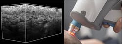
During the 2022 SPIE Biomedical Optics Symposium (BiOS), they outlined the development of a handheld in vivo probe incorporating dermoscopy and LC-OCT. And in a recent paper in Biomedical Optics Express, they discuss their development of paired ex vivo imagers that correlate those imaging modalities with Raman spectroscopy.
High-resolution depth imaging at speed
LC-OCT combines OCT with reflectance confocal microscopy (RCM). LC-OCT replaces the traditional point-illumination OCT beam with line illumination and replaces the conventional single detector with a line detector.
In traditional time-domain OCT, a low coherence source is directed to both a reference path and an object of interest. The reflected beams combine at a detector, which measures the intensity of the resultant interference. Short coherence length limits interference to a small thickness in the object. And the reference path length changes as the system records reflectance—an A-scan. Sweeping the OCT beam in a line creates a B-scan; stitched together, A-scans represent a vertical slice of the object.
With a cylindrical lens creating line illumination, LC-OCT creates an entire vertical slice with a single excursion of the reference path, parallelizing multiple A-scans to directly reconstruct a B-scan. Introducing a microscope objective increases the spatial resolution and allows the line detector to provide confocal filtering, rejecting light reflected from different planes, and creating a hybrid OCT/confocal microscope.
In both handheld in vivo and benchtop ex vivo incarnations, depth scanning is affected by moving the entire interferometer with a piezoelectric transducer, keeping the focal plane matched to the optical path difference.
The system creates horizontal images when a galvanometer mirror scans the illumination line across the target at a given depth. For this mode, the mirror at the end of the reference arm is sinusoidally modulated to acquire images at four different phases. These images are demodulated to reconstruct the reflected intensity, which enhances spatial resolution compared with a single reflectance image.
The system enables four imaging modes: vertical, horizontal, or combinations of both to create 3D reconstructions. In practice, the system combines horizontal images because it allows more control over volume reconstruction. It also presents a more direct path to future improvements in acquisition speed. The system currently acquires vertical and horizontal images at a live rate of 8 fps and can reconstruct 1.2 × 0.5 × 0.5 mm3 image volumes with nearly isotropic 1 µm resolution.
Putting skin in the game
These principles are applicable to different clinical targets. Here, the team aims at dermatology. Both the handheld and benchtop versions press a window against the skin, with index-matching oils maintaining focus and path length during acquisition. Currently, the researchers are investigating the clinical value added by Raman molecular fingerprinting.
Confocal Raman microspectroscopy (CRM) illuminates a specific point within tissue and directs wavelength-shifted inelastically scattered light to a spectrometer. Measuring the frequency shifts identifies molecules at the focal point. In this incarnation, a 785 nm laser source is fiber-coupled to a microscope objective, which gathers the scattered light and directs it to a fiber-coupled spectrometer. The microscope objective and fiber entrance provide confocal filtering.
Both the CRM and LC-OCT systems also incorporate a visible light camera for dermoscopy—images of the skin surface. The camera helps align the beams to surface features. But more precise registration is required to target the CRM to anatomical features revealed by LC-OCT.
The benchtop ex vivo LC-OCT identifies the coordinates of silicone and titanium dioxide beads in a calibration sample placed in a custom holder held in a magnetic kinematic mount. The sample holder is then transferred to an identical kinematic mount in the CRM system, where the strong Raman signal of the beads provides reference coordinates. Calculation of relative position and rotation allows the CRM to get within 20 µm of points of interest identified in an LC-OCT image.
The team demonstrated the performance by using CRM to distinguish five suspicious regions in a biopsy of tattooed skin, accurately distinguishing tattoo ink, inflammatory cells, and normal epidermal cells (see figure). Jonas Ogien, a research engineer with DAMAE Medical, summarizes the new capability: “This combination of morphological cellular-scale imaging, color surface imaging, and point molecular analysis opens up the possibility to perform 3D optical histology for all types of skin lesions.”
About the Author

Richard Gaughan
Contributing Writer, BioOptics World
Richard Gaughan is the Owner of Mountain Optical Systems and a contributing writer for BioOptics World.