Chromatic aberration aids in-depth imaging of biological tissue
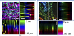
Researchers from the Beijing Advanced Innovation Center for Biomedical Engineering at the University of Beihang and collaborators developed a proof-of-principle chromatic confocal microscope with an extended depth of field, high resolution, and rapid image acquisition. Their instrument is capable of creating 3D images of biological tissue to depths of greater than 200 µm with a lateral resolution of 2.3 µm, filling the gap between optical coherence tomography and traditional confocal microscopy. With a two-dimensional 256 × 256-pixel lateral scan, the system can acquire a full volumetric image at a 1.5 Hz frame rate.
Challenges of living tissue
Cancer modifies the structure of individual cells and the tissue they compose. Clinical outcomes are generally much better if cancer is diagnosed at an early stage, so a method to detect small structural changes can provide great clinical value. But these structural changes are not limited to the tissue surface. For example, the prognosis for stomach cancer patients is improved if their gastric tumors can be resected, surgically removed, when they extend less than one-third the thickness of the submucosal layer, about 500 µm. So great clinical value would be provided by a method that can distinguish gastric tumors from normal tissue to those depths.
Chromatic confocal microscopy
The Beihang University team realized chromatic confocal microscopy offered a method of imaging tissue volumes without requiring mechanical axial scanning. This decreases exposure time and minimizes the complexity and physical volume of the imaging system.
But existing chromatic confocal systems didn’t offer a large enough focal shift to be effective for gastric tumor imaging. To solve this, the research team designed their own 11.5-mm-diameter, six-element miniature objective lens. The lens introduces a chromatic focal shift of 810 μm between light wavelengths of 650 nm to 950 nm, while having very low spherical aberration.
Light is delivered to the miniature objective through a 50,000-core optical fiber bundle. At the opposite, proximal end of the bundle, a filtered broadband light source sends 400–1000 nm light to a pair of steering mirrors, which direct the light to a single fiber core. Different wavelengths focus at different depths within the tissue and reflect back through the same fiber core. The reflected light from the proximal end of the fiber bundle is coupled to a single-mode fiber, which serves as the confocal spatial filter, rejecting light that doesn’t come from the focal plane of each wavelength.
This single-mode fiber delivers the light to a grating spectrometer with a 2048-pixel line-scan detector. Because the focal depth within tissue is a function of wavelength, the spectral distribution at the line-scan detector represents the depth distribution of reflective features within the tissue. As the steering mirrors scan across the field, they create a 2048-pixel spectral depth profile at each position within the field.
To verify the system’s performance and resolution, the team imaged a grooved silicon wafer, 10 µm aluminum beads in agarose gel, and a standard resolution target. The lateral resolution is 2.3 µm, and the average axial resolution is 12.2 μm in the range of 650–800 nm and 26.6 μm over wavelengths from 800 to 950 nm. The system is capable of completing a 256 × 256 raster scan at a 1.5 Hz rate. In the agarose gel phantom, the maximum imaging depth was 570 µm, sufficient for gastric tumor imaging.
Moving toward the clinic
The researchers imaged fresh gastric tissue excised from rats, as well as healthy and cancerous stomach tissue sections from humans. The chromatic confocal images show clear distinctions between dark cell nuclei and bright cell membranes with no labeling, which allows the researchers to distinguish the relatively regular cell morphology of normal tissue from the disordered structures within the tumor. It compares well with H&E histology images. In tissue, the system was able to create volumetric images to a depth of 220 µm.
Professor Pu Wang says their technology “will advance the diagnosis of many intra-organ diseases in locations such as the GI tract and bladder. It enables cellular-level resolution imaging of deep tissue with ultrafast 3D sectioning capability, which hasn’t been done in an endoscopy configuration.”
About the Author
Richard Gaughan
Contributing Writer, BioOptics World
Richard Gaughan is the Owner of Mountain Optical Systems and a contributing writer for BioOptics World.

