Aperiodic lensless imaging endoscope fiber reduces side lobes
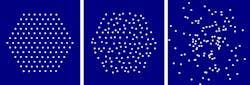
Lensless imaging endoscopes based on multicore optical fibers require no optomechanical elements on the end of the endoscope, allowing the instrument to be very small in diameter so that it can reach deep into tissue to image in a minimally invasive way. In addition, these types of endoscopes enable nonlinear imaging techniques such as two-photon excited fluorescence (TPEF) imaging. The endoscope is able to acquire an image without a lens because wavefront-shaping techniques allow the fiber output end to emit a single well-shaped wavefront, controlled from the other end of the fiber via a spatial light modulator to both scan and focus the output focal spot. In this way, 3D images are captured. However, because the fiber's multiple cores are conventionally in a periodic array, unwanted diffraction occurs, creating multiple side lobes around the focal spot.
Researchers from Aix-Marseille Université (Marseille, France), Université Lille (Villeneuve d'Ascq, France), and the Weizmann Institute of Science (Rehovot, Israel) have now fabricated and tested multicore fibers with aperiodic arrays that have enough pseudo-randomness to greatly reduce the number and intensity of side lobes, allowing for sharper imaging. For example, using a periodic array of 127 2-μm-diameter cores in a hexagonal array that fits within a multicore fiber diameter of 200 μm as a baseline core arrangement, the researchers compared it to two other core configurations: a pseudo-random arrangement with a pseudo-random parameter (PR) of 0.22 (which gives the hexagonal arrangement a jumbled appearance), and a random arrangement fitting within a 300 μm region. Calculations showed that the truly random arrangement reduced side lobes the most, with the pseudo-random arrangement also reducing lobes, but not as much. However, the truly random arrangement had some fiber cores too close to one another, causing undesired fiber coupling. Thus, the pseudo-random arrangement was most practical. A fabricated fiber proved the principle and was used to scan a small USAF chart. Reference: S. Sivankutty et al., arXiv:1606.08169v1 [physics.optics] (Jun. 27, 2016).
About the Author

John Wallace
Senior Technical Editor (1998-2022)
John Wallace was with Laser Focus World for nearly 25 years, retiring in late June 2022. He obtained a bachelor's degree in mechanical engineering and physics at Rutgers University and a master's in optical engineering at the University of Rochester. Before becoming an editor, John worked as an engineer at RCA, Exxon, Eastman Kodak, and GCA Corporation.