Laser light from the inside: fiber-optic nanotip electron gun enables easier nanoscale research
Fiber-optic nanotip electron sources are useful tools for materials science, bioimaging, and fundamental quantum research. In a conventional nanotip source, a metal nanotip is excited by external ultrafast laser pulses to produce free electrons; however, the need for external laser pulses makes the setup complicated and finicky.
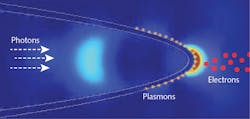
Now, scientists at the Department of Energy’s Oak Ridge National Laboratory (ORNL; Oak Ridge, TN) and the University of Nebraska (Lincoln, NE) have developed a fiber-optic nanotip electron source driven by laser pulses that arrive inside the fiber.1 As a result, the electron source is simple to set up and manipulate.
The researchers reported that firing intense laser pulses through a fiber-optic nanotip caused the tip to emit electrons, creating a fast “electron gun” that can be used to probe materials. The device allows researchers to quickly examine surfaces from any angle, which offers a huge advantage over less mobile existing techniques.
Since the mid-2000s, researchers have used sharp nanotips to emit electrons in tightly focused beams. The nanotips provide improved spatial and temporal resolution compared with other scanning electron microscopy techniques, helping researchers better track ongoing interactions at the nanoscale. In these techniques, electrons are emitted when photons excite the tips.
However, “previously, lasers had to track the tips, which is technologically a much harder thing to do,” says Herman Batelaan, who leads electron-control research at the University of Nebraska. The difficulty of the task limited how quickly images could be taken and from what position.
But Ali Passian of ORNL’s Quantum Information Science group had the idea that by firing laser light through a flexible optical fiber to illuminate its tapered, metal-coated nanotip from within, the result would be a more easily maneuverable tool. Passian teamed up with Batelaan and then graduate student Sam Keramati at the University of Nebraska. The Nebraska team used a femtosecond laser to shoot ultrashort, intense pulses through an optical fiber and into a vacuum chamber. In the chamber, the light moved through a gold-coated fiber nanotip that had been fabricated at ORNL. The team indeed observed controlled electron emission from the nanotip.
Analyzing the data, they proposed that the mechanism enabling the emission is not a simple one, but rather includes a combination of factors. One factor is that the shape and metal coating of the nanotip generates an electric field that helps push electrons out of the tip. Another factor is that this electric field at the nanotip’s apex can be enhanced by tuning the light to the tip's surface-plasmon-resonance wavelength.
An additional advantage of the new technique, they found, is that the fast-switching capacity of the laser source allows them to control electron emission at speeds faster than 1 ns—which will enable the capture of images at a very fast rate. Such images can then be pieced together to track complex interactions on the nanoscale.
Going CW
Pleased with these initial findings, the team decided to test if they could achieve a similar outcome with a far less powerful continuous-wave (CW) laser. To compensate for the lack of laser power, they upped the voltage at the nanotip, creating an energy potential difference they believed could help expel electrons. To their surprise, it worked.
“Now instead of having a powerful, extremely expensive laser, you can go with a $10 diode laser,” Batelaan notes.
Though CW lasers lack the fast switching capabilities of more powerful femtosecond lasers, slow switching offers its own advantages; namely, the possibility to better control the duration and number of electrons emitted by nanotips.
Electron ghost imaging
The team demonstrated, in fact, that the control provided by slow switching enabled electron emission within the bounds necessary for a new application called electron ghost imaging. Recently demonstrated light ghost imaging harnesses quantum properties of light to image sensitive samples, such as living biological cells, at very low exposure.
By bundling multiple fiber nanotips together, the team hopes to achieve electron ghost imaging on the nanoscale.
Source: https://www.ornl.gov/news/first-fiber-optic-nanotip-electron-gun-enables-easier-nanoscale-research
REFERENCE
1. Sam Keramati et al., New Journal of Physics (2020); https://doi.org/10.1088/1367-2630/aba85b.
About the Author
John Wallace
Senior Technical Editor (1998-2022)
John Wallace was with Laser Focus World for nearly 25 years, retiring in late June 2022. He obtained a bachelor's degree in mechanical engineering and physics at Rutgers University and a master's in optical engineering at the University of Rochester. Before becoming an editor, John worked as an engineer at RCA, Exxon, Eastman Kodak, and GCA Corporation.

