Optical Parametric Oscillators: Novel tunable lasers enable new nanoimaging techniques
Driven by the desire to characterize the electronic and vibronic properties of new materials with nanometer resolution, photonics researchers go through considerable effort to continuously refine nanoimaging techniques. Tip-enhanced Raman spectroscopy (TERS) is an approach that has been well recognized and relies on strongly localized enhancement of Raman scattering of laser light at the point of a near-atomically sharp tip. However, not least due to the lack of sources that would deliver laser light conveniently tunable throughout the visible spectral range, the vast majority of TERS experiments so far has been limited to single excitation wavelengths.
A recent study now demonstrates excitation-dependent hyperspectral imaging, exemplified on carbon nanotubes, by implementing a tunable continuous-wave optical parametric oscillator into a TERS setup. We take a closer look at the laser technology behind the experiment and illustrate the vast potential of the method.
Principles of optical parametric oscillators
Optical parametric oscillators (OPOs) might be considered as light sources that deliver coherent radiation very similar to lasers, but with two main differences between the devices.1 First, the OPO principle relies on a process referred to as parametric amplification in a nonlinear optical material, rather than on stimulated emission in a laser gain medium. Second, OPOs require a coherent source of radiation as a pump source, unlike lasers, which can be pumped with either incoherent light sources or sources other than light.
Figure 1 illustrates the basic scheme common to OPOs and other optical parametric devices. The process can be perceived as splitting of an incoming pump photon of high energy into two photons of lower energy, the latter usually referred to as signal and idler photons, respectively. It is essential to note that the overall process is subject to the conservation principles of photon energy and photon momentum (phase-matching condition), but otherwise does not have further fundamental restrictions, at least in theory. The huge potential of OPOs thus derives from their exceptional wavelength versatility, as they are in principle not limited by the wavelength coverage dictated by the energy levels and suitable transitions in a laser gain medium.
In practice, the OPO concept was experimentally demonstrated already more than half a century ago,2 but the progress in development and commercialization of turnkey devices has been stalled substantially by several technical obstacles.3 These obstacles have been easier to overcome at the high peak powers of pulsed devices, so that tunable OPOs operating in pulsed mode have become readily available from a variety of suppliers. Only relatively recently have there been comparable advances in continuous-wave (CW) OPO technology, which have spurred the development of commercial systems.
This progress has been mainly driven, on the one hand, by the increasing availability of cost-effective high-performance CW pump lasers and, on the other hand, by the advent and increasingly sophisticated design of new nonlinear crystals. As to pump lasers, the operation of CW OPOs puts stringent requirements on potential light sources in terms of preferential single-mode operation, noise characteristics, beam quality, and beam pointing stability.
Depending on power requirements of the end user, either high-performance diode-pumped solid-state (DPSS) lasers (for lower powers) or fiber-laser-based solutions (for higher powers) are typically used. As for nonlinear materials and novel crystal design techniques, it should be noted that the emergence of so-called quasi-phase-matched nonlinear materials like periodically poled lithium niobate (PPLN), whose crystal structure alternates with a certain periodicity, has been of great utility for the design of practical optical parametric devices.
Practical design considerations
While OPO technology appears to be ideally suited for generating tunable CW laser light across arbitrary wavelength ranges, one must keep in mind that the OPO process itself will always generate output at wavelengths that are longer than those used for pumping. Consequently, OPO devices operating across the visible spectral range either require UV pump sources or, alternatively, need to employ additional frequency conversion stages. As of now, only the latter approach has been proven to be technically practicable and operationally stable in commercial turnkey systems.
The essential building blocks of a tunable CW OPO designed to cover the visible range are shown in Figure 2.4 The operational principle relies on a cascaded sequence of nonlinear optical processes within two cavities, referred to as OPO and SHG cavities, respectively. As outlined above, pump laser photons are first split into pairs of photons of lower energy (signal and idler). The particular OPO scheme used is commonly referred to as singly resonant OPO cavity design: For a certain operational wavelength of the entire system, the cavity is operated on resonance at either a particular signal wavelength or a particular idler wavelength. Therefore, a precisely movable stack of periodically poled nonlinear crystals allows for broad wavelength coverage. At a particular wavelength selection, a crystal layer with a suitable poling is automatically selected and its poling period fine-adjusted through a temperature-control loop. At the same time, the effective OPO cavity length is actively stabilized to a multiple integer of the selected operational wavelength. While circulating one of the generated (signal or idler) waves resonantly inside the OPO cavity, its counterpart can be extracted for wavelength conversion into the visible spectral range by another nonlinear process.As shown in Figure 2, this wavelength conversion takes place in a second, separate cavity by frequency doubling of the primary OPO cavity output, a process widely known as second-harmonic generation (SHG). Though this configuration is technically practicable and provides favorable operational stability, it should be mentioned that alternative designs, like intracavity frequency doubling, have been successfully demonstrated in the lab.
Raman spectroscopy of carbon nanotubes
How does widely tunable laser light, as can be provided by the sources described above, advance nanoimaging techniques? To answer this question, carbon nanotubes (CNTs) have been selected in a recent study as a testbed for proof of principle of a novel experimental approach based on TERS.5 We recall that a CNT is essentially a strip of graphene (a one-atom-thick sheet of carbon) rolled up into a cylinder along a so-called chiral vector with two indices (n,m). It is this chiral vector that completely determines the CNT microscopic structure—that is, its tube diameter and the chiral angle along the tube axis.
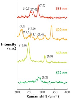
Raman scattering has been well established as one of the main techniques to identify the chiral vectors of CNTs experimentally.6 So-called radial breathing modes (RBMs) that correspond to collective movements of carbon atoms in the radial direction serve as fingerprints of particular (n,m) configurations in the Raman spectrum.
The usual CNT Raman scattering signals are typically very weak, and therefore of little practical relevance. However, the Raman scattering efficiency is significantly enlarged if the laser energy matches the energy of an optically allowed electronic transition—an enhancement process referred to as resonance Raman scattering.6 In other words, for a particular laser excitation wavelength, the observed Raman signals (respective radial breathing modes) from a mixture of CNTs will derive only from those CNTs that are in electronic resonance with the laser excitation (see Fig. 3). Note that the Raman data, recorded for a mixture of CNTs in solution, is to be perceived as a compositional analysis, but does not contain any spatial information whatsoever.Excitation-tunable tip-enhanced Raman spectroscopy
The three main components of a TERS setup include: A laser light source for excitation, an atomic-force microscope (AFM) equipped with a sharp metallic tip, and a Raman spectrometer recording the inelastically scattered radiation.5 The basic physical principle behind TERS relies on so-called localized surface plasmons that are excited by the laser light in the microscope tip. These plasmons generate a strongly localized electromagnetic field, which not only enhances the incoming and Raman-scattered radiation by orders of magnitude, but also ensures a highly localized excitation of the sample under study. Thus, by recording tip-enhanced Raman spectra intensities as a function of the tip position, TERS allows for nanoimaging with a spatial resolution down to below 10 nm.
Figure 4 illustrates the sequence of events and results of a TERS experiment carried out at a single laser excitation wavelength (633 nm) on a film of a CNT mixture.5 In a first step, a so-called composed Raman spectrum is recorded by placing the microscope tip at a particular x, y position in close proximity to the CNT film. The resulting composed Raman spectrum encompasses the radial breathing mode peaks of several CNTs (all of them in electronic resonance to the excitation wavelength). In a second step, the microscope tip is retracted and the far-field spectrum recorded without the tip-enhanced Raman contribution to the signal. By subtracting the far-field spectrum from the composed spectrum, the pure tip-enhanced Raman spectrum is obtained. Eventually, from the pure tip-enhanced Raman spectrum, the tube species underneath the tip position can be unambiguously identified—a CNT with (7,5) chirality in the example shown in Figure 4a.For a spatial image of the particular CNT, the outlined procedure is repeated: The tip position is scanned stepwise over the sample surface and at each point the intensity of the pure tip-enhanced Raman peak determined. Figure 4b shows the result of such a scan and images the position of a (7,5) CNT in a 550 × 140 nm2 area. As can be seen, the CNT is around 800 nm long and bent in a steplike shape.
The full beauty of the experimental approach now unfolds when realizing that the imaging capability of the setup is no longer limited to a subset of CNTs that happen to be in electronic resonance to a particular excitation wavelength, as has been the case for the vast majority of TERS experiments. On the contrary, the examination of the sample under study can be in principle performed for a quasi-continuum of wavelengths that is covered by the tunable laser light source.
In the present example, this allows unprecedented access to a broad variety of CNT species—Figure 5 shows excitation-tunable tip-enhanced Raman spectroscopy (e-TERS) methodology recently reported.5 By using four different excitation wavelengths, it becomes possible to identify and image a total of nine different CNT species within one and the same sample area. We point out that the e-TERS nanoimages in Figure 5 visualize, for the first time, the shape and orientation of different CNT species in spatially overlapping arrangements. And they are no longer limited to an observation window that is dictated by a narrow band of electronic transitions that fall to be in resonance with a single excitation wavelength.Outlook
The experimental demonstration of excitation-tunable tip-enhanced Raman spectroscopy comes in tandem with the availability of novel tunable laser light sources based on OPO technology. From the general laser technology point of view, the performance characteristics of OPOs make them competitive alternatives to conventional lasers and related technologies for the generation of widely tunable CW radiation. From the experimental methodology point of view, we expect e-TERS to open new experimental horizons for studying the electronic and vibronic properties of matter on the nanometer scale—it is tantalizing to envision the application of this method to the existing broad variety of 1D and 2D materials.
ACKNOWLEDGEMENTS
The authors gratefully acknowledge enlightening discussions and support by Stephanie Reich and her group at the Free University Berlin.
REFERENCES
1. R. Paschotta, Optical Parametric Oscillators, Encyclopedia of Laser Physics and Technology Ed. 1, Wiley-VCH (2008).
2. J. A. Giordmaine and R. C. Mills, Phys. Rev. Lett., 14, 973 (1965).
3. M. Ebrahim-Zadeh, Optical Parametric Oscillators, in Handbook of Optics Ed. 2, McGraw-Hill, Ed. 2 (2001).
4. J. Sperling and K. Hens, Optik & Photonik, 13, 22 (2018).
5. N. S. Mueller, S. Juergensen, K. Höflich, S. Reich, and P. Kusch, “Excitation-tunable tip-enhanced Raman spectroscopy,” J. Phys. Chem C, accepted.
6. C. Thomsen and S. Reich, Raman Scattering in Carbon Nanotubes, Top. Appl. Phys., 108, 115 (2007).
About the Author
Korbinian Hens
Product Manager, HÜBNER Photonics
Korbinian Hens is product manager at HÜBNER Photonics, Kassel, Germany.
Patryk Kusch
Postdoctoral Researcher, Freie Universität Berlin
Patryk Kusch is a postdoctoral researcher at Freie Universität Berlin in Germany.
Jaroslaw Sperling
Business Developer, Femtosecond Fiber Lasers at Menlo Systems
Jaroslaw Sperling is business developer for femtosecond fiber lasers at Menlo Systems (Martinsried, Germany). With a background in ultrafast laser spectroscopy, he holds a Ph.D. in physical chemistry from the University of Vienna (Austria).

![FIGURE 2. The beam path inside a commercial CW OPO system [4] is shown here in a schematic. In the first step, a 532 nm laser pumps a nonlinear crystal to generate signal and idler photons (in a 900–1300 nm range). Wavelength selection and subsequent second-harmonic generation (SHG) converts either signal or idler photons into the visible range of the spectrum (450–650 nm). The green arrow depicts the pump laser beam; dark red and light red arrows depict the signal and the idler beam (arbitrary assignment). FIGURE 2. The beam path inside a commercial CW OPO system [4] is shown here in a schematic. In the first step, a 532 nm laser pumps a nonlinear crystal to generate signal and idler photons (in a 900–1300 nm range). Wavelength selection and subsequent second-harmonic generation (SHG) converts either signal or idler photons into the visible range of the spectrum (450–650 nm). The green arrow depicts the pump laser beam; dark red and light red arrows depict the signal and the idler beam (arbitrary assignment).](https://img.laserfocusworld.com/files/base/ebm/lfw/image/2019/01/1901lfw_spe_f1.png?auto=format,compress&fit=max&q=45?w=250&width=250)


![FIGURE 5. Nanoimages of carbon nanotubes are recorded with four different excitation wavelengths [5]. The different tube species are labeled by their chiral indices in the nanoimages. FIGURE 5. Nanoimages of carbon nanotubes are recorded with four different excitation wavelengths [5]. The different tube species are labeled by their chiral indices in the nanoimages.](https://img.laserfocusworld.com/files/base/ebm/lfw/image/2019/01/1901lfw_spe_f5.png?auto=format,compress&fit=max&q=45?w=250&width=250)