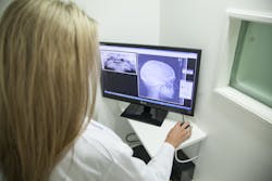
Image processing algorithm helps make x-rays for children safer
Photonics scientists at the Warsaw University of Technology (WUT; Warsaw, Poland), working in collaboration with innovation incubator ACTPHAST 4.0 and medical imaging company Italray (Scandicci, Florence, Italy), have developed a new algorithm that ‘autocorrects’ unclear, low-dose digital x-rays to generate a higher-contrast image, meaning young children can receive safer x-ray scans.
When having an x-ray or computed tomography (CT) scan, beams enter the body and ricochet around inside—or become ‘scattered.’ Given that the scatter signal interferes with the primary contrast of the patient’s physical features such as bones or organs, this scattering process creates ‘noise’ and leads to a loss in image quality, making resulting x-rays appear blurred.
The scatter, however, can be counteracted with an ‘antiscatter grid’—a metal plate made of lead strips to encourage parallel beams—improving the image contrast. But, this grid normally requires a higher dose of x-rays, and can therefore be dangerous to small children.
X-rays can emit harmful ionizing radiation—high-energy particles that penetrate tissue to reveal internal organs and bone structures—which can damage DNA. While scientists have often sought to reduce this ionizing radiation, traditionally it has come at the expense of the type of detector and image resolution. Recognizing this, the team of scientists has been able to address the image quality problem as a result of scatter from the perspective of the acquired data and the digital image processing.
The research team’s method works by minimizing the scattering process by taking the original image and estimation of the scatter signal, explains Wojciech Krauze, the team’s project manager. By partially ‘reversing’ the scatter, the digital image processing algorithm is able to reduce the amount of noise signal, essentially ‘autocorrecting’ the blurred image instantly, he says.
“The result is a ‘scatter grid quality’ image without the need for an actual anti scatter grid,” Krauze says.
