Smartphone-based 'dermascopes' image skin cancer at low cost
A team of University of Arizona (Tucson, AZ) researchers has demonstrated that standard smartphone technology can be adapted to image skin lesions, providing a low-cost, accessible medical diagnostic tool for skin cancer.
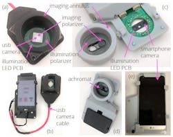
In their paper that describes the work, the researchers developed two dermascopes using a smartphone-based camera and a USB-based camera. Both dermascopes integrate LED-based polarized white-light imaging (PWLI), polarized multispectral imaging (PMSI), and image-processing algorithms, and were able to map and delineate between the dermal chromophores indicative of melanoma, and the general skin redness known as erythema.
Leveraging a smartphone camera to image skin lesions improves both the efficiency and efficacy of skin-lesion diagnostics, says Brian W. Pogue, SPIE Fellow and MacLean Professor of Engineering at Dartmouth College (Hanover, NH), who serves as the Editor-in-Chief of the Journal of Biomedical Optics, the journal in which the paper appears. “The functionality and performance for the detection of skin cancers is very important,” he says. “The cellphone camera approaches proposed and tested in this paper introduce key steps in a design that will provide simple, accurate diagnoses. While there are always ways to make medical imaging systems work, they can often be too expensive to be a viable commercial success. Similarly, devices can be made so complex that they aren’t readily adopted. Here, the already-familiar platform is both low-cost and intuitive to use. The simplicity of the approach here successfully combines the need for simple diagnostic tools with high precision. This is the kind of innovation that will potentially allow for easier adoption in clinical use.”
Full details of the work appear in the Journal of Biomedical Optics.
