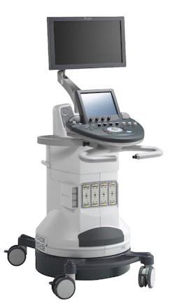
Multimodal imaging system could enable biopsy-free breast screening
An international team of scientists from the Horizon2020 project SOLUS has developed a noninvasive multimodal imaging system that uses ultrasound and light technologies to easily differentiate between benign or malignant breast lesions, eliminating the need to perform a biopsy. To perform the screening, a clinician scans the breast with a handheld ‘smart optode’ pen probe that combines light and sound to collate blood parameters and tissue constituents.
Using a technique called diffuse optical imaging, a method that has already provided breakthroughs in neuroscience, wound monitoring, and cancer detection, scientists can monitor changes in concentrations of oxygenated and deoxygenated hemoglobin, collagen, lipids, and water present in a suspected tumor against a preprogrammed set of results.
While mammography is accurate in detecting breast lesions, many women encounter false-positive results—that is, a positive detection of a lump, but with no malignant cancer present. Clinicians, however, do not necessarily know whether a lesion is cancerous or harmless and have to resort to invasive procedures, such as biopsies, to make an accurate diagnosis.
Able to provide a malignant or benign diagnosis, the SOLUS scanner reads a number of different parameters to create a thorough characterization of tissue. Gathering the total blood volume and oxygenation, collagen, water and lipid content, together with stiffness and morphologic information, the system produces an accurate, while-you-wait diagnosis.
Aiming for 95% sensitivity and 90% specificity, the project has combined commercial ultrasound imaging and elastography with novel diffuse optical imaging approaches.
Harnessing photonics and acoustics, the system gathers diagnostic information from the composition of tissue and blood by sending pulses of infrared light and ultrasound a few centimeters into the breast tissue. A clinician can then determine whether a lump is benign or malignant given that cancerous tissue is characterized by high hemoglobin and water content, and low lipid content.
Having undergone extensive laboratory trials, the SOLUS team plans to validate the system in real clinical settings at the end of 2020 and into 2021, says Paola Taroni from the Politecnico di Milano in Italy, the SOLUS project coordinator.
The SOLUS project includes nine partners from European countries. It is supported by the European Commission in the framework of the EU Horizon 2020 ICT program with a grant of €3.8 million under the Photonics Public Private Partnership.
For more information, please visit photonics21.org.
