Laser-based x-ray images bone sample in 3D, within minutes
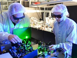
Researchers from Ludwig-Maximilians University (LMU), the Max Planck Institute of Quantum Optics (MPQ), and the Technical University of Munich (TUM; all in Munich, Germany) have demonstrated that a laser-based x-ray source enables tomographic reconstruction of the three-dimensional (3D) fine structure of a bone sample within a few minutes rather than several hours. The breakthrough was made possible by the further development of ATLAS, the high-performance laser in LMU's Laboratory for Extreme Photonics (LEX Photonics) at LMU on the Research Campus in Garching, Germany. Reconstruction of the sample from the imaging data was also facilitated using specially designed computer programs.
Related: A new era of bioimaging with x-rays
To harness the advantages of synchrotron radiation for general use in medicine, the research team has been exploring the application of high-performance lasers to the production of x-rays. In their setup, hydrogen atoms are irradiated with extremely intense pulses of laser light. The highly energetic optical fields strip the electrons from the atoms and part of the ionized plasma electrons are accelerated. Simultaneously, these electrons oscillate in the plasma fields, which causes them to emit the desired synchrotron radiation—that is, high-intensity x-rays. Moreover, this whole process takes place over a path-length of <15 mm. So, laser-based x-ray sources have a far smaller footprint, and are much less expensive to build, than conventional synchrotrons, but produce x-radiation of comparable quality.
In early trials carried out at the Max Planck Institute in 2015, the research team was able to derive the 3D structure of an insect from 2D projection images taken from different angles. For the latest experiments, performed at LEX Photonics, Stefan Karsch and his colleagues have boosted the pulse rate, photon yield, and photon energies, and this time they chose to image a sample of human bone. Thanks to an improved processing algorithm developed by Franz Pfeiffer and his group at TUM, the team needed to collect significantly less data than before. Accordingly, the complete tomogram could be obtained within less than three minutes.
The project was conceived and initiated in the Munich-Centre for Advanced Photonics (a Cluster of Excellence), and is undergoing further development at the Center for Advanced Laser Applications (CALA; also in Garching). The laser systems available at CALA are expected to significantly enhance the efficiency of the source and the quality of the radiation generated, thus making this new form of tomography available for clinical applications.
Details of the team's work appear in the journals Optica and Nuclear Instruments and Methods A.