Portable camera images the retina without need for dilation, and is low-cost
A team of researchers from the University of Illinois at Chicago (UIC) College of Medicine (Chicago, IL) and Massachusetts Eye and Ear/Harvard Medical School (Boston, MA) has developed a low-cost (less than $200), portable camera that can image the retina without the need for pupil-dilating eye drops.
Related: Retinal imaging advances research, disease diagnosis
Bailey Shen, a second-year ophthalmology resident at the UIC College of Medicine, explains that there are often times when dilating patients is not an option, such as in neurosurgery patients. "Also, there are times when we find something abnormal in the back of the eye, but it is not practical to wheel the patient all the way over to the outpatient eye clinic just for a photograph," he adds.
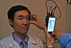
The prototype camera, which is pocket-sized, can take pictures of the back of the eye without eye drops, Shen says. The pictures can be shared with other doctors, or attached to the patient’s medical record.
The camera is based on the Raspberry Pi 2 computer, a low-cost, single-board computer designed to teach children how to build and program computers. The board hooks up to a small, low-cost infrared (IR) camera, and a dual IR- and white-light-emitting diode (LED). The camera also includes a lens, a small display screen, and several cables.
The camera works by first emitting IR light, which the iris (the muscle that controls the opening of the pupil) does not react to. Most retina cameras use white light, which is why pupil-dilating eye drops are needed. The IR light is used to focus the camera on the retina, which can take a few seconds. Once focused, a quick flash of white light is delivered as the picture is taken. Cameras exist that use this same IR/white light technique, but they are bulky and often cost thousands of dollars.
Shen's camera photos show the retina and its blood supply as well as the portion of the optic nerve that leads into the retina. It can reveal health issues that include diabetes, glaucoma, and elevated pressure around the brain.
Full details of the work appear in the Journal of Ophthalmology; for more information, please visit https://doi.org/10.1155/2017/4526243.
