Photoacoustic endoscope shows promise for Crohn’s disease treatment
Researchers from the University of Michigan (Ann Arbor, MI) have developed an endoscope that could improve the view of intestinal changes caused by Crohn's disease, a painful and debilitating form of inflammatory bowel disease. The endoscope is used for photoacoustic imaging, a bioimaging method that uses light to produce sound waves in tissue that can be captured with ultrasound imaging.
In Crohn's disease, both inflammation and fibrosis cause the development of strictures (areas of narrowing) in the intestines. Although strictures caused by inflammation can be treated with drugs, the ones caused by fibrosis must be removed surgically. "The difficulty in accurately assessing the presence and development of fibrosis in the strictures adds a great deal of complexity to Crohn's disease management decisions," says Guan Xu, leader of the research team.
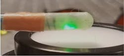
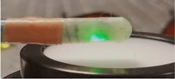
Researchers developed an endoscope that can perform photoacoustic imaging; the new device could give doctors a better view of intestinal changes caused by Crohn's disease. (Image credit: Guan Xu, University of Michigan)
In their study, the researchers developed a capsule-shaped photoacoustic endoscope to examine whether this imaging technique could be used to characterize inflammation and fibrosis in intestinal strictures. The capsule-shaped probe was 7 mm in diameter and 19 mm long.
They designed the endoscope to deliver near-infrared light at 1310 nm because this wavelength is absorbed by collagen protein, which is characteristic of fibrosis. The light absorption causes the protein to expand slightly, leading to a mechanical vibration that can be captured using ultrasound imaging. To generate a strong signal, the researchers constructed the endoscope to maximize delivery of 1310 nm light.
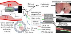
The researchers are working to miniaturize the endoscope so that it could be used in the working channel of a colonoscope; this would allow a surgeon to view photoacoustic images prior to performing treatment. (Image credit: Guan Xu, University of Michigan)
The researchers tested their new endoscope in rabbit models with intestinal narrowing caused by either inflammation only or a mix of fibrosis and inflammation. The experiments showed the endoscopic photoacoustic imaging approach could quantitatively differentiate inflammatory from fibrotic intestinal strictures. Another study in rabbits demonstrated that the endoscope could also quantify the development of fibrosis over time.
The researchers are now working to make the endoscope small enough to pass through the instrument channel of a colonoscope, a flexible fiber-optic instrument used to examine the large intestine. This could provide a surgeon with diagnostic information immediately before treatment without the need for additional procedures.
Full details of the work appear in the journal Biomedical Optics Express.


