Prototype portable neuroimaging system to study stroke recovery, epilepsy
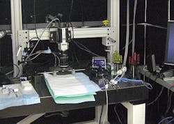
Researchers at the University of Toronto in Canada have developed a portable neuroimaging prototype for studying the effects of stroke recovery and epilepsy. Equipped with an electron-multiplying charge-coupled device (EMCCD) camera, the system continuously monitors blood flow to the brain in non-anesthetized animals.
University of Toronto Assistant Professor Ofer Levi and colleagues demonstrated how they were able to simultaneously measure two modalities—blood oxygenation and flow speeds—in one system. The system also allowed them to statistically distinguish between veins and arteries, something never before accomplished. Other scaled-down imaging solutions are limited to tracking a single modality or require the use of multiple complex systems.
The team plans to create a smaller, portable version of the system that will allow them to image fully conscious, active animals participating in normal behaviors, says Levi. Doing so will enable long-term chronic monitoring of epilepsy dynamics and recovery from stroke in preclinical animal studies—which could lead to an instrument that can be used for clinical use in patients, he says.
Levi simultaneously quantified flow changes in blood vessels and measured dynamics in oxygenation to track neural activity in real time using an EMCCD camera (QImaging's Rolera EM-C2). The camera's low read noise allowed him to rapidly detect low-light signals with high sensitivity. So, he and his team were able to show dynamics of flow and oxygenation changes in response to induced ischemia in a wide field of view with speeds that at times exceeded 100 fps, making it possible to calibrate the entire brain flow map, he says.
For more information on the team's work, which has been published in Biomedical Optics Express, please visit http://www.opticsinfobase.org/boe/abstract.cfm?URI=boe-3-4-777.