'3D photografting' method uses laser beams to precisely attach molecules
Researchers at the Vienna University of Technology (Vienna, Austria) have developed a method that uses a laser beam to fix biological molecules at exactly the right position in a 3D material, and on a micrometer scale. The work is promising for artificially growing biological tissue or for creating biosensors.
Dubbed "3D photografting," the method begins with a hydrogelâa type of scaffold made of macromolecules arranged in a loose meshwork. Between those molecules, there are large pores through which other molecules or cells can migrate.
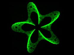
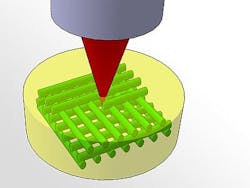
Then, specially selected molecules are introduced into the hydrogel meshwork and a laser beam irradiates certain points. At the positions where the focused laser beam is most intense, a photochemically labile bond is broken. That way, highly reactive intermediates are created, which locally attach to the hydrogel quickly. The precision depends on the laserâs lens system; the researchers' system was able to obtain 4 µm resolution.
The researchers' method can be used to artificially grow biological tissue: Like a climbing plant clinging to a trellis, cells need a scaffold to which they attach. In natural tissue, the extracellular matrix accomplishes this by using specific amino acid sequences to signal to the cells where they are supposed to grow. In the lab, scientists are trying to use similar chemical signals. Cell attachment could be guided on 2D surfaces, but in order to grow larger tissues with a specific inner structure (such as capillaries), a 3D technique is required.
Depending on the application, different molecules can be used. 3D photografting is not only useful for biomedical engineering, but also for photovoltaics or sensor technology.
The researchers' work has been published in Advanced Functional Materials; for more information, please visit http://onlinelibrary.wiley.com/doi/10.1002/adfm.201290098/abstract.
-----
Follow us on Twitter, 'like' us on Facebook, and join our group on LinkedIn
Laser Focus World has gone mobile: Get all of the mobile-friendly options here.
Subscribe now to BioOptics World magazine; it's free!


