Photoacoustic imaging tool could detect and diagnose cervical cancer noninvasively
A team of researchers at Central South University (Changsha, Hunan, China) has demonstrated that photoacoustic imaging, which is already under investigation for detecting skin or breast cancers and for monitoring therapy, also has the potential to be a new, faster, cheaper, and noninvasive method to detect, diagnose, and stage cervical cancer with high accuracy.
Related: Photoacoustic tomography is ready to revolutionize
In their study, the researchers examined 30 cervical tissue samples in vitro using photoacoustic imaging. Some were taken from healthy women and some of the samples contained the cancerous cells of a cervical tumor. They found that photoacoustic imaging could distinguish the cancerous from the normal tissue and had the potential to evaluate the stage of the cancer.
"Our results show that the photoacoustic imaging may have great potential in the clinical diagnosis of cervical cancer," says Jiaying Xiao, an assistant professor of biomedical engineering at Central South University, who led the research. "The technique is noninvasive and can detect the lesions in the cervical canal, an area conventional methods fail to observe. The photoacoustic imaging can also evaluate the invasion depth of cervical lesions more effectively."
According to Xiao, one of the conventional screening methods is colposcopy, a technique in which doctors use a special magnifying device to examine the vulva, vagina, and cervix and identify suspicious lesions, which are then biopsied. Although colposcopy is effective in detecting lesions around the external cervical orifice and cervical biopsy is considered the gold standard for diagnosing cancer in the cervix, these conventional methods are both time- and labor-intensive and expensive. Moreover, colposcopy may miss some cancer lesions.
For example, Xiao explains, a considerable number of the cervical cancer cases start from lesions in the cervical canal, but the interior of cervical canal cannot be directly observed with colposcopy. Because of that, these lesions may not be detected until the cancer has advanced and spread to other tissues. So Xiao says that there is still a need to develop new diagnostic technologies with higher accuracy, deeper penetration, larger scanning regions, and lower cost.
According to Xiao, photoacoustic imaging combines the high optical contrast of pure optical imaging with the high spatial resolution and the deep imaging depth of ultrasound. In the technique, short laser pulses are employed to irradiate biological tissues. Some of the laser energy is absorbed by the tissues and converted into heat, leading to rapid thermal expansion inside the tissues that produces ultrasonic waves. The generated waves are then detected by an ultrasonic sensor to form photoacoustic images of the tissues.
By using hemoglobin as the contrast agent or indicator, photoacoustic imaging is highly sensitive to abnormal angiogenesis, the abnormal formation of new blood vessels, which is a hallmark of the cancer tumors. By irradiating a tissue with a short laser pulse, the part with abnormal angiogenesis will absorb more laser energy than the surrounding tissues and produce a different acoustical signature. Using ultrasound detectors, researchers can map these photoacoustic signals and identify the locations of lesions.
In the study, Xiao and his colleagues conducted 30 in vitro experiments with tissue samples representing different cancer stages. In each experiment, researchers embedded one piece of normal cervical tissue and one piece of cervical lesion from the same person in a cylindrical phantom for simultaneous photoacoustic imaging. Part of each sample was also sent to histological evaluation for cross-photoacoustic imaging.
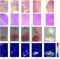
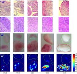
By processing all of the photoacoustic imaging data with computer programs, scientists obtain a 2D en face photoacoustic image called a depth maximum amplitude projection (DMAP) image, which shows the optical absorption distribution or photoacoustic contrast of the sample. The image can help scientists identify the lesion location and evaluate different cancer stages of cervical cancer.
“Stronger absorption from the cervical lesions is observed compared to that of normal tissue,” Xiao says. Statistical results also show that the mean optical absorption (MOA) of the cervical lesions is closely related to the severity of cervical cancer.
Compared with other cervical screening methods, Xiao adds, photoacoustic imaging also has a much higher penetration depth by using ultrasound as the localizing signal, which is less scattered than light. Therefore, the optical absorption distribution can be obtained deep in the biological tissue.
While the work is preliminary, it provides solid experimental basis for future studies, showing for the first time that photoacoustic imaging has the potential to make better diagnoses and help save lives, Xiao says. The next step, he adds, is to prove the applicability of photoacoustic imaging in tumor models and ultimately in patients.
“Our ultimate goal is to develop an endoscopic photoacoustic imaging probe scanning the cervical canal, which would be a quicker, cheaper, and noninvasive method for the diagnosis of cervical cancer,” Xiao says.
Full details of the work appear in the journal Biomedical Optics Express; for more information, please visit http://dx.doi.org/10.1364/BOE.6.000135.
-----
Follow us on Twitter, 'like' us on Facebook, connect with us on Google+, and join our group on LinkedIn
Subscribe now to BioOptics World magazine; it's free!

