Optical method can detect proteins by their scattered light
Researchers at the Max Planck Institute for the Science of Light (Erlangen, Germany) have developed an optical method that enables direct detection of the scattered light of individual proteins via their shadows to not only make biomedical diagnoses more sensitive, but also provide new insights into fundamental biological processes. The work could someday lead to earlier disease diagnosis and more effective treatment.
Related: Light-scattering approach reveals nano-scale cellular changes for early cancer detection
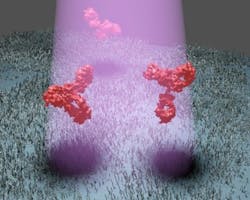
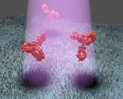
Vahid Sandoghdar, director at the Max Planck Institute for the Science of Light, and Marek Piliarik, a postdoc in Sandoghdar's division, are now able to produce a much clearer image without the need for elaborate attachment of luminous markers to the target proteins. Their method, dubbed interferometric detection of scattering (iSCAT), involves shining laser light onto a microscope slide on which the relevant proteins have been captured. The proteins then scatter the laser light, thus casting a weak shadow.
"Until now, it was thought that if you want to detect scattered light from nanoparticles, you have to eliminate all background light," explains Sandoghdar. "However, in recent years we've realized that it is more advantageous to illuminate the sample strongly and visualize the feeble signal of a tiny nanoparticle as a shadow against the intense background light." By doing so, this allows the background light to interfere with the weak scattered light so that the desired signal is amplified.
The researchers have also learned to eliminate noise by taking a snapshot with the iSCAT microscope not only after they have dripped a solution containing the desired protein onto the sample holder, but also beforehand. "Since most of the optical noise generated by nanoscopic irregularities of the sample do not change, we can subtract one image from the other and thus eliminate the noise," says Piliarik. The target proteins then stand out clearly from the background as dark spots, even though the shadow of the protein is only one ten-thousandth or even one hundred-thousandth as dark as the background.
Piliarik and Sandoghdar are able to detect various proteins as shadows under the microscope not only in pure solutions, but also in mixtures containing concentrations of other proteins that are up to 2000 times greater. "The specificity of the detection method is not limited by the optics, but by the selectivity of the substances used to bind the target proteins to the microscope slide," says Piliarik. As the researchers found, the amount of contrast between the protein and background depends on the size of the target particle. They even found indications that this contrast can reveal information about the shape of the protein.
"The strength of our method lies not only in the fact that it is so sensitive and that we can count target proteins in a sample," says Sandoghdar. "iSCAT also shows us the exact position of particles." Having knowledge of protein interactions is crucial for understanding diseases and a broad range of biological processes. In the future, iSCAT might be possible for observing particles even smaller than proteins, such as RNA fragments, to watch how they interact with other substances. To this end, the researchers are working to further improve the signal-to-noise ratio.
Full details of the work appear in the journal Nature Communications; for more information, please visit http://dx.doi.org/10.1038/ncomms5495.
-----
Follow us on Twitter, 'like' us on Facebook, and join our group on LinkedIn
Subscribe now to BioOptics World magazine; it's free!

