MOLECULAR IMAGING/ONCOLOGY: Non-toxic nanoparticle opens new options for tumor imaging, treatment
A fluorescing, non-toxic and biodegradable organic nanoparticle with a versatile structure may enable a new approach to the treatment of various types of cancer and to drug delivery. That’s because of the way in which it uses light and heat, say the Canadian researchers who developed the innovation at Princess Margaret Hospital (PMH, Toronto, Ontario).
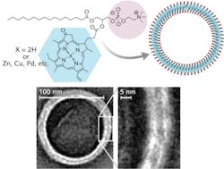
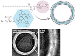
“We combined two naturally occurring molecules (chlorophyll and lipid) to create a unique nanoparticle that shows promise for numerous diverse light-based (biophotonic) applications,” says Dr. Gang Zheng, senior scientist, Ontario Cancer Institute (OCI), PMH. The nanostructure accumulates in tumors and absorbs light efficiently, and can be filled with drugs to treat the targeted tissue.
“The nanoparticle can also be used for photoacoustic imaging, which combines light and sound to produce a very high-resolution image that can be used to find and target tumors,” says OCI doctoral student Jonathan Lovell, who co-authored a paper describing the work.1 He adds that once the nanoparticle hits its target, it fluoresces.
“There are many nanoparticles out there, but this one is the complete package, a kind of one-stop shopping for various types of cancer imaging and treatment options that can now be mixed and matched in ways previously unimaginable,” says Zheng, noting that its “unprecedented safety” in the body is the icing on the cake.
1. J. Lovell et al., Nature Materials 10, 324–332 (2011).
More BioOptics World Current Issue Articles
More BioOptics World Archives Issue Articles