Label-free, pulsed laser-driven imaging tool tracks nanotubes in cells, blood
Carbon nanotubes have potential applications in drug delivery to treat diseases and imaging for cancer research, but up until now there has been no technique to see both metallic and semiconducting nanotubes in living cells and the bloodstream, according to Ji-Xin Cheng, associate professor of biomedical engineering and chemistry at Purdue University (West Lafayette, IN).
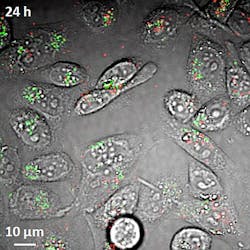
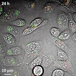
To tackle this problem, Cheng's team developed a new imaging tool that tracks carbon nanotubes in living cells and the bloodstream, which could help to boost their use in biomedical research and clinical medicine. The imaging technique, called transient absorption, uses a pulsing near-infrared laser to deposit energy into the nanotubes, which then are probed by a second near-infrared laser. As a bonus, transient absorption is also label-free.
The nanotubes have a ~1 nm diameter, which makes them far too small to be seen with a conventional light microscope. One challenge in using the transient absorption imaging system for living cells was to eliminate the interference caused by the background glow of red blood cells, which is brighter than the nanotubes.
The team solved this problem by separating the signals from red blood cells and nanotubes in two separate "channels." Light from the red blood cells is slightly delayed compared to light emitted by the nanotubes. The two types of signals are "phase-separated" by restricting them to different channels based on this delay.
The researchers used the technique to see nanotubes circulating in the blood vessels of mice earlobes, enabling them to visualize the nanotubes circulating in the bloodstream in real time. This is important for drug delivery in order to know how long nanotubes remain in blood vessels after they are injected, says Cheng.
The structures, called single-wall carbon nanotubes, are formed by rolling up a one-atom-thick layer of graphite called graphene. The nanotubes are inherently hydrophobic, so some of the nanotubes used in the study were coated with DNA to make them water-soluble, which is required for them to be transported in the bloodstream and into cells.
The researchers also have taken images of nanotubes in the liver and other organs to study their distribution in mice, and they are using the imaging technique to study other nanomaterials such as graphene.
The National Science Foundation supported the team's work, which has been published online in Nature Nanotechnology. For more information, please visit http://www.nature.com/nnano/journal/vaop/ncurrent/full/nnano.2011.210.html.
-----
Follow us on Twitter, 'like' us on Facebook, and join our group on LinkedIn
Follow OptoIQ on your iPhone; download the free app here.
Subscribe now to BioOptics World magazine; it's free!

