Adaptive optics visualizes, characterizes vascular disease early and noninvasively
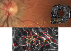
Researchers over time have found that abnormalities in the retinal vasculature correlate with a diverse range of conditions, including diabetes, hypertension, stroke, Alzheimer's disease, migraine, and glaucoma. The earliest anomalies in vascular disease arise in the microvasculature and current imaging methods lack the spatiotemporal resolution to track blood flow at the capillary level.
Recognizing this, researchers at the University of Melbourne in Australia have demonstrated direct, noninvasive optical imaging of erythrocyte flow in human retinal capillaries, and validated its use as an investigative tool for early signs of disease. Led by Phillip Bedggood and Andrew Metha, the group modified an ophthalmoscope with adaptive optics to achieve 1.5 µm spatial resolution and a flood illumination system. They also replaced their systems's previous CCD camera with a 5.5 Mpixel scientific CMOS (sCMOS) camera (Andor Technology's Neo sCMOS camera), which enabled data capture at up to 460 fps. When tested on the eye of a human subject, the range of erythrocyte velocities was found to be in agreement with previously published figures based on retinal leukocytes.

"Phillip Bedggood's work is highly significant," says Orla Hanrahan, application specialist at Andor. "In directly visualizing red blood cells in the retinal capillaries, they can now use this approach to study the dynamics of the retinal vascular supply under normal conditions and in response to functional stimulation of the retina. More importantly, they will be able to investigate how the dynamics change in the presence of cerebrovascular and cardiovascular diseases, such as diabetes, stroke, and Alzheimer's."
For more information on the team's work, please visit http://www.optometry.unimelb.edu.au/research/methaLab.html.
-----
Follow us on Twitter, 'like' us on Facebook, and join our group on LinkedIn
Subscribe now to BioOptics World magazine; it's free!