Clinical research project evaluates noninvasive IR imaging of burn wounds
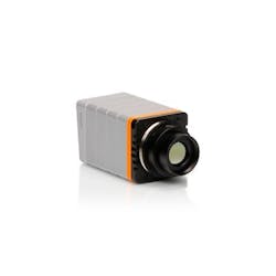
Infrared (IR) imagers and cameras maker Xenics (Leuven, Belgium) is collaborating with the VU University Medical Center (VUmc; Amsterdam, Netherlands) in groundbreaking clinical research programs, including a project focused on burn wounds in collaboration with the Dutch Burn Center (Beverwijk, Netherlands). Central to the setup at VUmc is Xenics' Gobi-640 thermal camera for noninvasive diagnostics and treatment evaluation.
Assessing burn wounds in terms of incurred tissue damage and their healing prospects requires effective diagnostic tools to determine whether the probability of scar development would necessitate a skin transplantation. Such diagnostic and treatment procedures should be based on a simple, compact, and cost-effective apparatus suited for the clinical environment for an easy, reliable, and straightforward interpretation and statistical analysis. They also should allow real-time monitoring of the healing progress over the duration of the treatment.
Now that IR cameras have become easy to use without complicated calibration and cooling measures, Xenics' Gobi-640 thermal imaging camera is proving suitable for medical research and clinical practice due to its compact plug-and-play layout, on-board processing capabilities, and extensive software platform for analysis. According to Rudolf M. Verdaasdonk, Ph.D., a professor at VU and Head of the Department of Physics and Medical Technology at the VU Medical Center, the camera can observe physiological processes in burn wounds based on perfusion—the blood streaming through the peripheral vessels towards damaged skin tissue and showing the presence of an intact or damaged microvasculature, depending on the temperature measured.
A specific advantage of thermography in the clinical study carried out at the Dutch Burn Center, under supervision of plastic surgeon professor Paul van Zuijlen, is that it enables the continued observation and real-time evaluations of burn wounds to notice improvements in the healing process.
Verdaasdonk says that thermography could provide much better diagnostic options in the early stages of burn wounds, and it could enable a much better evaluation of the healing progress. "I foresee that in the future, every medical practitioner will have some smartphone device with a thermal camera in their pocket. This could even extend to popular health apps used at home."
-----
Follow us on Twitter, 'like' us on Facebook, connect with us on Google+, and join our group on LinkedIn
Subscribe now to BioOptics World magazine; it's free!
