X-ray laser maps key signaling protein, with potential for better drug development
An international team of researchers, led by the Van Andel Institute (Grand Rapids, MI), performed an x-ray laser experiment at the Department of Energy's SLAC National Accelerator Laboratory (Menlo Park, CA) to reveal never-before-seen details of the human body's cellular switchboard that regulates sensory and hormonal response. The work could enable more highly targeted and effective drug development with fewer side effects to treat conditions including high blood pressure, diabetes, depression, and some types of cancer.
Related: X-ray laser can solve protein structures from scratch
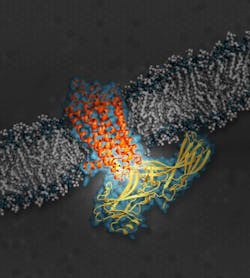
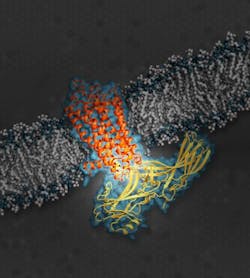
Ultrabright x-rays using SLAC’s Linac Coherent Light Source (LCLS) enabled the research team to complete the first 3D atomic-scale map of a key signaling protein called arrestin while it was docked with a cell receptor involved in vision. The receptor is a well-studied example from a family of hundreds of G protein-coupled receptors (GPCRs), which are targeted by about 40 percent of drugs on the market. Its structure, while coupled with arrestin, provides new insight into the on/off signaling pathways of GPCRs.
Arrestins and another class of specialized signaling proteins called G proteins take turns docking with GPCRs. Both play critical roles in the body’s communications “switchboard,” sending signals that the receptors translate into cell instructions. These instructions are responsible for a range of physiological functions.
Before the study at SLAC, little was known about how arrestins—which serve a critical role as the “off” switch in cell signaling, opposite the “on” switch of G proteins—dock with GPCRs, and how this differs from G protein docking. The latest research helps scientists understand how a docked arrestin can block a G protein from docking at the same time, and vice versa. Many of the available drugs that activate or deactivate GPCRs block both G proteins and arrestins from docking.
“The new paradigm in drug discovery is that you want to find this selective pathway—how to activate either the arrestin pathway or the G-protein pathway but not both—for a better effect,” says Eric Xu, a scientist at the Van Andel Institute who led the experiment. The study notes that a wide range of drugs would likely be more effective and have fewer side effects with this selective activation.
Xu said he first learned about the benefits of using SLAC’s x-ray laser for protein studies in 2012. The microscopic arrestin-GPCR crystals, which his team had painstakingly produced over years, proved too difficult to study at even the most advanced type of synchrotron, a more conventional x-ray source.
In the LCLS experiments, Xu’s team used samples of a form of human rhodopsin—a GPCR found in the retina whose dysfunction can cause night blindness—fused to a type of mouse arrestin that is nearly identical to human arrestin. Measuring thousandths of a millimeter, the crystals—which had been formed in a toothpaste-like solution—were oozed into the x-ray pulses at LCLS, producing patterns that when combined and analyzed allowed researchers to reconstruct a complete 3D map of the protein complex.
Xu says his group hopes to conduct follow-up studies at LCLS with samples of GPCRs bound to different types of signaling proteins.
Full details of the work appear in the journal Nature; for more information, please visit http://dx.doi.org/10.1038/nature14656.
Follow us on Twitter, 'like' us on Facebook, connect with us on Google+, and join our group on LinkedIn

