New microendoscope doubles sensitivity of esophageal cancer screenings
A low-cost, portable, battery-powered microendoscope developed by Rice University (Houston, TX) bioengineers could eventually eliminate the need for costly biopsies for many patients undergoing standard endoscopic screening for esophageal cancer.
Related: Tiny OCT endoscope could have big impact on esophageal cancer precursor Barrett's
A clinical study involving 147 U.S. and Chinese patients undergoing examination for potentially malignant squamous cell tumors explored whether the low-cost, high-resolution fiber-optic imaging system could reduce the need for unnecessary biopsies when used in combination with a conventional endoscope.
In the study, all 147 patients with suspect lesions were examined with both a traditional endoscope and the new microendoscope. Biopsies were obtained based upon the results of the traditional endoscopic exam. A pathology exam revealed that more than half of those receiving biopsies—58 percent—did not have high-grade precancer or cancer. The researchers found that the microendoscopic exam could have spared unnecessary biopsies for about 90 percent of the patients with benign lesions.
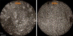
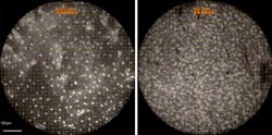
To determine whether a biopsy is needed for a histological exam, health professionals often use endoscopes, small cameras mounted on flexible tubes that can be inserted into the body to visually examine an organ or tissue without surgery. The new high-resolution microendoscope uses a 1-mm-wide fiber-optic cable that is attached to the standard endoscope. The cable transmits images to a high-powered fluorescence microscope, and the endoscopist uses a tablet computer to view the microscope’s output. The microendoscope provides images with similar resolution to traditional histology and allows endoscopists to see individual cells and cell nuclei in lesions suspected of being cancerous. By providing real-time histological data to endoscopists, the microendoscope can help rule out malignancy in cases that would otherwise require a biopsy.
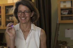
The lab that developed the microendoscope is led by Rebecca Richards-Kortum, Rice University's Stanley C. Moore Professor of Bioengineering, professor of electrical and computer engineering, and director of Rice 360°: Institute for Global Health Technologies. Richards-Kortum's lab specializes in the development of low-cost optical imaging and spectroscopy tools to detect cancer and infectious disease at the point of care. Her research group is particularly interested in developing technology for low-resource settings, and the microendoscope was developed as part of that effort. It is battery-operated, inexpensive to operate, and requires very little training. Results from the clinical study verified that both experienced and novice endoscopists could use the microendoscope to make accurate assessments of the need for a biopsy.
Clinical studies of the microendoscope are either planned or underway for a dozen types of cancer, including cervical, bladder, oral, and colon cancers.
Full details of the work appear in the journal Gastroenterology; for more information, please visit http://dx.doi.org/10.1053/j.gastro.2015.04.055.
Follow us on Twitter, 'like' us on Facebook, connect with us on Google+, and join our group on LinkedIn


