Quantum dot, activated by light, reveals how protein corona forms

Using a cadmium selenide (CnSe) quantum dot (quantum dots are used in in vivo imaging of tumor cells, detection of biomolecules, and measurement of pH changes, among other bioimaging applications), researchers at Syracuse University in New York collaborated to understand how the protein corona forms and what is different about the quantum dot before and after the formation of the corona.
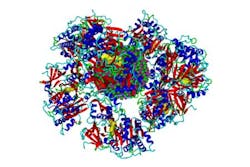
When quantum dots are introduced in biological media, proteins surround the nanoparticles and form a corona. The formation of the protein corona changes the sensitivity of the quantum dots to light.
Professor Shikha Nangia of Syracuse's Department of Biomedical and Chemical Engineering and Professor Ari Chakraborty of the Department of Chemistry have developed a novel, multilevel computational approach. This method combines the strengths of quantum mechanics, molecular mechanics, classical molecular dynamics, and Monte Carlo techniques. Because of this work, it is now possible to perform computer simulation of protein-quantum dot complexes that were previously considered to be beyond the scope of computational investigations. Now that this methodology has been created, it can be applied to bigger and more complex quantum dot systems.
Full details of the work appear in the Journal of Chemical Theory and Computation; for more information, please visit http://dx.doi.org/10.1021/ct500681m.
-----
Follow us on Twitter, 'like' us on Facebook, connect with us on Google+, and join our group on LinkedIn
Subscribe now to BioOptics World magazine; it's free!