Ultrafast laser light enables mini x-ray source for 3D imaging of tissue
A team of researchers from Ludwig-Maximilians University, the Max Planck Institute of Quantum Optics, and Technical University of Munich (TUM; all in Munich, Germany) have developed a method that uses ultrafast laser-generated x-rays and phase-contrast x-ray tomography to produce three-dimensional (3D) images of soft tissue structures in organisms.
Related: X-ray laser maps key signaling protein, with potential for better drug development
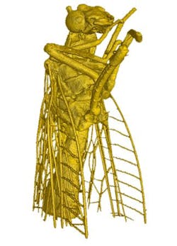
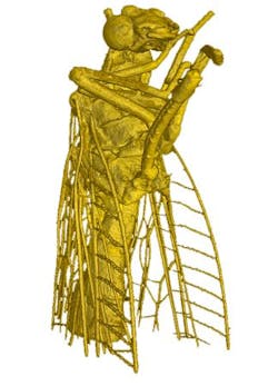
With laser light, the researchers from the Laboratory of Attosecond Physics of the Max Planck Institute of Quantum Optics and TUM built a miniature x-ray source to capture 3D images of ultrafine structures in the body of a living organism for the first time. Using light-generated radiation combined with phase-contrast x-ray tomography, the scientists visualized ultrafine details of a fly measuring a few millimeters. Until now, such radiation could only be produced in expensive ring accelerators measuring several kilometers in diameter. By contrast, the laser-driven system in combination with phase-contrast x-ray tomography only requires a university laboratory to view soft tissues. The new imaging method could make future medical applications more cost-effective and space-efficient than is possible with today’s technologies.
When physicists Prof. Stefan Karsch and Prof. Franz Pfeiffer illuminated a tiny fly with x-rays, the resulting image captured the finest hairs on the wings of the insect. The scientists coupled their technique for generating x-rays from laser pulses with phase-contrast x-ray tomography to visualize tissues in organisms. The result is a 3D view of the insect in unprecedented detail.
The x-rays required were generated by electrons that were accelerated to nearly the speed of light over a distance of approximately 1 cm by laser pulses lasting around 25 fs. The laser pulses have a power of approximately 80 TW (80 × 1012 W).
The technology is particularly interesting for medical applications, as it is able to distinguish between differences in tissue density. Cancer tissue, for example, is less dense than healthy tissue. The method therefore opens up the prospect of detecting tumors that are less than 1 mm in diameter in an early stage of growth before they spread through the body and exert their lethal effect. For this purpose, however, researchers must shorten the wavelength of the x-rays even further to penetrate thicker tissue layers.
Full details of the work appear in the journal Nature Communications; for more information, please visit http://dx.doi.org/10.1038/ncomms8568.
Follow us on Twitter, 'like' us on Facebook, connect with us on Google+, and join our group on LinkedIn

