PHOTOACOUSTICS/PRECISION SURGERY: Lens transforms pulsed light into precision sound scalpel
State-of-the-art ultrasound technology can focus sound waves tightly enough to generate heat, enabling doctors to pulverize kidney stones and prostate tumors. But the focal spot is typically a few centimeters wide—too bulky to treat delicate vasculature, for instance. Now, a lens that converts pulsed laser light to high-pressure sound waves "can enhance the focal accuracy 100-fold," says Hyoung Won Baac. Developed at the University of Michigan (U-M; Ann Arbor, MI), the optical approach generates high-frequency (>15 MHz), high-amplitude focused ultrasound, and promises a new tool for high-precision noninvasive surgery.
Baac, a research fellow at Harvard Medical School, worked on this project as a U-M doctoral student in the lab of Jay Guo, co-author of a paper describing the technique.1 The optoacoustic approach that converts light to high-amplitude sound waves is not new, but until now the sound signal hasn't been strong enough to be useful.
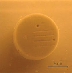
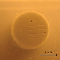
To amplify the signal, the researchers coated their lens with layers of carbon nanotubes and polydimethylsiloxane. The carbon nanotube layer absorbs the light and generates heat, expanding the polydimethylsiloxane, which boosts the signal dramatically by rapid thermal expansion. The resulting high-frequency sound waves work in tissues by creating shockwaves and microbubbles. Thus, treatment is enabled by pressure, not heat.
The researchers were able to concentrate sound waves to just 75 × 400 μm. In experiments, they accurately detached a single ovarian cancer cell and drilled a 150 μm hole in an artificial kidney stone in less than a minute. Guo thinks it might be able to operate painlessly because the finely focused beam could avoid nerves.
1. H. W. Baac et al., Sci. Rep., 2, 989, doi:10.1038/srep00989 (2012).

