BIOMEDICAL IMAGING/X-RAY: Lasers replace accelerator, magnets in device that makes synchrotron x-rays widely accessible
Synchrotron x-rays boost the quality of medical imaging and lower doses of radiation compared to x-ray devices currently used in hospitals worldwide. But synchotron x-ray devices have traditionally been too large and expensive to be widely accessible. Now, researchers at the University of Nebraska-Lincoln's (UNL) Extreme Light Laboratory have developed a new way to generate these x-rays using a powerful, compact laser.1
Physics professor Donald Umstadter, director of the Extreme Light Laboratory, led the research project. He compares the synchrotron x-ray breakthrough to the development of personal computers, giving more people access to power once available only via large and costly mainframes. Shrinking components of advanced laser-based technology will increase the feasibility of producing high-quality x-rays in medical and university research laboratories, which in turn could lead to new applications for the x-rays, he says.
Because the new x-ray device could fit in a hospital or truck, it could facilitate widespread applications for advanced x-ray technology, including the ability to find cancerous tumors at earlier stages or study extremely fast reactions that occur too rapidly for observation with conventional x-rays.
In traditional synchrotron machines, electrons are accelerated to extremely high energy and then made to change direction periodically, leading them to emit energy at x-ray wavelength. Magnets change the direction of the electrons and produce x-rays.
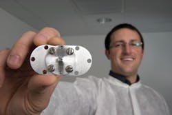
The UNL team replaced both the electron accelerator and the magnets with laser light. They first focused their laser beam onto a gas jet, creating a beam of relativistic electrons. They then focused another laser beam onto the accelerated electron beam. This rapidly vibrated the electrons, causing them to emit a bright burst of synchrotron x-rays—a process referred to as Compton scattering. Remarkably, the light's photon energy was increased during this process by a million-fold. And yet, the combined length of the accelerator and synchrotron was less than the size of a dime.
"The x-rays that were previously generated with compact lasers lacked several of the distinguishing characteristics of synchrotron light, such as a relatively pure and tunable color spectrum," Umstadter says. "Instead, those x-rays resembled the 'white light' emitted by the sun." The new laser-driven device produces x-rays over a much larger range of photon energies, extending to the energy of nuclear gamma rays. Even fewer conventional synchrotron x-ray sources are capable of producing such high photon energy.
Key to the breakthrough was finding a way to collide the two micro-thin beams—the scattering laser beam and the laser-accelerated electron beam.
1. N. D. Powers et al., Nat. Photon., 8, 28–31 (2014); doi:10.1038/nphoton.2013.314.