IN VIVO IMAGING: Optical molecular imaging: closer to clinical
Optical molecular imaging continues its steady march toward the clinic as demonstrated by a number of talks at the World Molecular Imaging Conference (September 23–26, Montreal, QC, Canada). At the meeting’s kick-off keynote, Nobel Prize winner Roger Tsien stressed that surgery and radiation therapy offer key opportunities for optical molecular imaging. His group has developed synthetic molecules called activatable cell penetrating peptides (ACPP) that selectively accumulate in diseased tissue. Brighter and smaller than fluorescent proteins, these molecules help visualize small metastases in the lung and are useful for surgical guidance.
“We have found that molecular fluorescence image guidance with ACPP is superior to standard surgery,” said Tsien. The addition of ACPP to a tumor site gives the surgeon a roadmap to fine structures such as nerves. Using optical molecular probes like ACPP could help surgeons more aggressively go after tumors because they can easily visualize the nerves woven through a solid tumor.
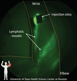
The use of the near-infrared fluorophore indocyanine green (ICG) in microdoses has led to the first images of the human lymphatic system as described by John Rasmussen from The Brown Foundation Institute of Molecular Medicine, University of Texas Health Science Center (Houston, TX) (see Fig. 1). The effective use of small amounts of ICG may allow for more rapid approval by the U.S. Food and Drug Administration (FDA) thus enabling more widespread clinical use. Often called the forgotten circulation system of the body, the lymph system flushes and filters waste from the blood and transports proteins through the body. When this system fails, chronic swelling and reduced immune response occur.
Novel applications
The meeting highlighted several emerging optical technologies and applications. For instance, Adam de la Zerda of Stanford University (Stanford, CA) described a prototype photoacoustic system that images new blood vessel growth in the eye. Early detection of such growth could lead to better treatments for wet macular degeneration and diabetic retinopathy.
Gordon Turner of the Novartis Institutes for Biomedical Research (Cambridge, MA) reported the use of optical projection tomography to study drug effects on heart disease. Using optical imaging provides faster, more robust data than traditional whole slide imaging.
And Ning Zhang of Caliper Life Sciences (Hopkinton, MA) discussed how bioluminescence imaging permits longitudinal studies to monitor the survival of adipose tissue grafts. This work is particularly important for cosmetic and reconstructive surgery because adipose tissue is used in breast augmentation, reconstruction of breast defects, and wound healing.
New agents, new systems
According to exhibitors, optical imaging agents and multimodality systems will drive the field of molecular imaging in the next few years. While small animal preclinical studies will continue to provide a solid base for the optical molecular imaging market, optical imaging agents ready for clinical trials could play a key role in wider acceptance of optical techniques for clinical applications.
One such case is Li-Cor Biosciences’s (Lincoln, NE) IRDye 800CW PEG contrast agent. The agent has shown promising results imaging the human lymphatic system, and according to company representatives will be in clinical trials within a year. A number of companies such as Caliper Life Sciences, VisEn Medical (Bedford, MA), and Care-stream Molecular Imaging (Rochester, NY) are ramping up production of current imaging agent products and spending more time and money developing new agents in wavelength ranges applicable to humans.
As the agents find new applications, new systems will follow. On display at the meeting were several systems either in clinical use or ready for use. They included ART/Advanced Research Technologies Inc.’s (Montreal, QC, Canada) Softscan breast imager, Fluoptics’s (Grenoble, France) handheld real-time imager for oncology surgery, and O2View’s (Marken, the Netherlands) real-time Artemis, a multi-spectral NIR fluorescence camera system for surgical applications.
In the area of preclinical optical imaging, the hardware has reached a level where few technical challenges remain. To enhance their products, then, companies are upgrading image analysis software and combining distinct imaging modalities into a single system. A number of new or improved systems were introduced at WMIC, many with multimodal capabilities. Caliper introduced the IVIS Lumina XR which combines both optical imaging and anatomical information from x-ray. VisEn, which produces systems for fluorescence molecular tomography (FMT), introduced the FMT 1500 for small to mid-sized facilities, and an updated version of its FMT 2500 (see Fig. 2). The next-generation system, the LX, operates at four different wavelengths and comes with adapters that allow for CT, MRI, and PET scanning. ART/Advanced Research Technologies Inc. introduced Optix MX3, a whole animal 3-D in vivo imager able to co-register optical information with data from CT scans.Market “speeding up”
Although many sectors of the economy have taken hits this year, the molecular imaging market doesn’t appear to have lost much ground. Caliper President Kevin Hrusovsky described optical molecular imaging as a “vibrant field” and noted that sales on Caliper’s IVIS system have topped $50 million. Three years ago sales were $30 million.
“Each year going forward will represent five years. Things are speeding up,” said Hrusovsky.
About the Author


