OPTOGENETICS/SUBSTANCE ABUSE: Laser brain stimulation reduces alcohol consumption in rats
Low-frequency light activation of dopamine (DA) neurons reduces a drinking binge by 54% and significantly delays the start of drinking. These are the findings of research on rats to elucidate the mechanisms that drive alcohol abuse.1
Despite the prevalence of alcohol in our society, little is known about the neurochemical alterations that drive abuse. Research has pointed to DA as the primary neurotransmitter involved in positive reward; the ventral tegmental region (VTA) is an important region of the DA system that is connected by dopaminergic neurons to another brain region, the nucleus accumbens (NA). This area plays an integral role in the regulation of motivated behaviors, and alterations of this circuit have long been hypothesized to contribute to the cause of alcoholism.
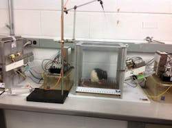
Many studies have implicated a DA mechanism in the regulation of ethanol drinking. However, it is not possible to selectively stimulate DA release from discrete DA neurons using electrical or chemical stimulation. But such stimulation is necessary for testing a causal link between DA neuron activation and drinking. Optogenetics allowed the scientists—from Wake Forest University and Wake Forest School of Medicine (both in Winston-Salem, NC), SUNY University at Buffalo (Buffalo, NY), and the University of North Carolina at Chapel Hill—to stimulate dopaminergic cells with exceptional selectivity and temporal precision. They demonstrated that only specific pulses (5 Hz frequency and 50 s duration) of 473 nm light with a maximum power output of 100 mW affected drinking behavior.
While several transgenic strategies have been developed to target opsins (light-sensitive proteins) to dopaminergic neurons in rodents, the research team developed a novel viral approach that can be used in any mammalian species and potentially even humans. Here, the expression of channelrhodopsin-2 (ChR2), a mutated cell membrane protein that opens ion channels in response to light, was driven by a tyrosine hydroxylase (TH) promoter. The TH promoter confined production of ChR2 to only DA neurons, and not any other type of neurons; laser light was then applied to the VTA region to stimulate DA transmission.
The researchers showed that they could deliver the ChR2 gene to DA neurons and selectively control the timing/amount of DA released in the NA without affecting DA dynamics in neighboring brain regions.
1. C. E. Bass et al., Front. Behav. Neurosci., 7, 173 (2013); doi:10.3389/fnbeh.2013.00173.