Blue light-activated neurons from stem cells could restore function to paralyzed muscles
Scientists at University College London (UCL) and King’s College London, both in England, have found a new way to artificially control muscles using light, with potential to restore function to muscles paralyzed by conditions such as motor neuron disease and spinal cord injury.
Related: LCD projector light stimulates neurons and muscles of moving worms
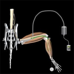
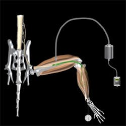
The technique involves transplanting specially designed motor neurons created from stem cells into injured nerve branches. These motor neurons are designed to react to pulses of blue light, allowing for fine-tuning of muscle control by adjusting the intensity, duration, and frequency of the light pulses.
Related: Blue light prompts protein clustering and, in turn, advances optogenetics
The research team demonstrated the method in mice, in which the nerves that supply muscles in the hind legs were injured. They showed that the transplanted stem cell-derived motor neurons grew along the injured nerves to connect successfully with the paralyzed muscles, which could then be controlled by pulses of blue light.
“Following the new procedure, we saw previously paralysed leg muscles start to function,” says Professor Linda Greensmith of the MRC Centre for Neuromuscular Diseases at UCL’s Institute of Neurology, who co-led the study. “This strategy has significant advantages over existing techniques that use electricity to stimulate nerves, which can be painful and often results in rapid muscle fatigue. Moreover, if the existing motor neurons are lost due to injury or disease, electrical stimulation of nerves is rendered useless as these too are lost.”
Muscles are normally controlled by motor neurons, which are specialized nerve cells within the brain and spinal cord. These neurons relay signals from the brain to muscles to bring about motor functions such as walking, standing, and even breathing. However, motor neurons can become damaged in motor neuron disease or following spinal cord injuries, causing permanent loss of muscle function and resulting in paralysis.
“This new technique represents a means to restore the function of specific muscles following paralysing neurological injuries or disease,” explains Professor Greensmith. “Within the next five years or so, we hope to undertake the steps that are necessary to take this ground-breaking approach into human trials, potentially to develop treatments for patients with motor neuron disease, many of whom eventually lose the ability to breathe, as their diaphragm muscles gradually become paralyzed. We eventually hope to use our method to create a sort of optical pacemaker for the diaphragm to keep these patients breathing.”
The light-responsive motor neurons that made the technique possible were created from stem cells by Dr. Ivo Lieberam of the MRC Centre for Developmental Neurobiology at King’s College London.
“We custom-tailored embryonic stem cells so that motor neurons derived from them can function as part of the muscle pacemaker device,” says Lieberam, who co-led the study. “First, we equipped the cells with a molecular light sensor. This enables us to control motor neurons with blue light flashes. We then built a survival gene into them, which helps the stem-cell motor neurons to stay alive when they are transplanted inside the injured nerve and allows them to grow to connect to muscle.”
Full details of the work appear in the journal Science; for more information, please visit http://dx.doi.org/10.1126/science.1248523.
-----
Follow us on Twitter, 'like' us on Facebook, and join our group on LinkedIn
Subscribe now to BioOptics World magazine; it's free!

