Hat-like molecular cages allow light activation of signaling peptides in vivo
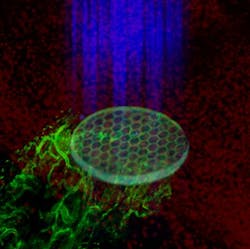
To transport biomaterials that contain peptide-signaling molecules into living animals, Georgia Institute of Technology (Georgia Tech; Atlanta, GA) researchers and colleagues used hat-like molecular cages to cover binding sites on the peptides that are normally recognized by cell receptors. When activated by ultraviolet (UV) light, the cages detach and reveal the peptides in vivo. If their method could be made to work in humans, it could help provide more precise timing for processes essential to regenerative medicine, cancer treatment, immunology, stem cell growth, and other areas.
Related: Light now able to activate cell surface receptors, with promising implications
“Many biological processes involve complex cascades of reactions in which the timing must be very tightly controlled,” says Andrés García, a Regents Professor in the George W. Woodruff School of Mechanical Engineering at Georgia Tech and principal investigator for the project. “Until now, we haven’t had control over the sequence of events in the response to implanted materials. But with this technique, we can deliver a drug or particle with its signal in the ‘off’ position, then use light to turn the signal ‘on’ precisely when needed.”
During the five-year project, the research team—which included Ted Lee and Jose Garcia from Georgia Tech and Aranzazu del Campo from the Max Planck Institute—modified peptides that normally trigger cell adhesion to present the molecular cage to disguise them. They showed that disguised peptides introduced into animal models on biomaterials could trigger cell adhesion, inflammation, fibrous encapsulation, and vascularization responses when activated by light. They also showed that the location and timing of activation could be controlled inside the animal by simply shining light through the skin.
The work involved numerous controls to ensure that the triggering observed by the researchers was actually done by exposure of the peptides—not the light, or the removal of the protective cage. The researchers also had to demonstrate that the “hats” were stable enough that they didn’t come off spontaneously, but only when the link between the molecular cage and the peptide was severed by the UV light.
Among the experiments was use of the peptide to attract cells that would attach themselves to the biomaterial. “We showed that if we left the hat on, there would be few cells attracted to the material, García says. “But when we take the hat off, we recruited a lot of cells to the material. That shows we can activate the peptide, and that the activation has a biological consequence.”
Another experiment showed that the timing of peptide activation could affect the quantity of fibrosis, an immune system response that builds a protective capsule around an implanted biomaterial. By delaying the exposure of the peptides until after the bulk of the inflammation reaction had taken place, the thickness of the fibrosis capsule was significantly reduced, allowing it to be better incorporated into the body.
In another experiment, the researchers showed that removing the hats could trigger the growth of blood vessels into the material. This vascularization is critical in regenerative medicine, but must take place at the right time to be successful.
“We showed that if you keep the hat on, you get no vessel in-growth into the material,” explains García. “But if we turn on the light, we get growth of new blood vessels into the material. We can control what happens and when it happens by when we expose the protective cages to light.”
In the future, photochemists at the Max-Planck Institute will be working on alternative cages that would be triggered by different wavelengths of light. As much as 90 percent of the UV light used in the experiments was lost in passing through the skin of the animal model, limiting the use of that wavelength to locations immediately below the skin.
Development of alternate molecular cages could allow sequential activation by light, and light activation of molecules at locations deeper inside the body.
Full details of the work appear in the journal Nature Materials; for more information, please visit http://dx.doi.org/10.1038/nmat4157.
-----
Follow us on Twitter, 'like' us on Facebook, connect with us on Google+, and join our group on LinkedIn
Subscribe now to BioOptics World magazine; it's free!


