Optical techniques help assess the impact of toxic agents in cells
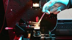
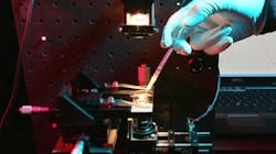
Seeking a way to accurately determine the toxicity of nanomaterials in cells, researchers at École Polytechnique Fédérale de Lausanne (EPFL; Lausanne, Switzerland) turned to optical techniquesâincluding measuring the light absorbed by certain proteinsâto measure the oxidative stress that some of them provoke on cells.
Related: Raman, nanotechnology approach detects, tracks and kills cancer cells
Related: Low-intensity laser enables label-free approach to computed 3D live cell imaging
When a cell is exposed to a toxic product or a pathogen, this causes the internal equilibrium between the oxidants and antioxidants within the cell to break. Then the former, generally oxygen derivatives, are produced in excessive quantities and start to attack the cellâs proteins, sugars, and its membrane. This brings about a faster cellular aging, causes certain diseases to the cell, and may even lead to its death.
Thus, the overproduction of such oxidants is a sign that the cell is stressed, and that is exactly what researchers wanted to measure. At the same time, they noticed that cytochrome c, a protein present in the cellular membrane, was a particularly interesting biosensor. They found that when it was exposed to certain wavelengths of light, this protein would absorb less light when in the presence of one of these oxidizing agents: hydrogen peroxide. Consequently, they developed a complex method for measuring the variations of light absorbed by cytochrome c. Finally, they tested and verified their method on small unicellular algae.
To this day, there were no truly reliable methods for measuring oxidative stress continuously and without damaging the cells. This new test has opened interesting possibilities for identifying not only the effect of nanomaterials, but also, on a wider perspective, the way cells react to an external perturbation. In addition, during their experiments researchers were able to observe that the toxicity of certain products could be conditioned and modulated by its surrounding environment. For example; a nanomaterial may be less dangerous under a laboratory microscope than within a riverâs waters.
"The test that we propose is highly sensitive and able to indicate the concentration of oxygen derivatives in a thorough and detailed way," says Olivier Martin, director of the Nanophotonics and Metrology Laboratory (NAM) at EPFL. "Since it is based in assessing a substance released outside the cells, it is also noninvasive. Therefore, it does not destroy the living organism and can be applied over a period of several hours making it possible to observe the evolution of the situation over time."
Full details of the work appear in the journal Scientific Reports; for more information, please visit http://www.nature.com/srep/2013/131209/srep03447/full/srep03447.html.
-----
Follow us on Twitter, 'like' us on Facebook, and join our group on LinkedIn
Subscribe now to BioOptics World magazine; it's free!

