X-ray laser experiment could help in new drug development for brain disorders

A team of scientists at Stanford University (California), using an x-ray laser, have revealed never-before-seen details of how the brain sends rapid-fire messages between its cells. They mapped the 3D atomic structure of a two-part protein complex—neuronal SNAREs and synaptotagmin-1—that controls the release of signaling chemicals called neurotransmitters from brain cells. Understanding how cells release those signals in less than one-thousandth of a second could help launch a new wave of drug development for treating brain disorders.
Related: X-ray laser maps key signaling protein, with potential for better drug development
The experiments, conducted at the Linac Coherent Light Source (LCLS) x-ray laser at the Department of Energy’s SLAC National Accelerator Laboratory, build upon decades of previous research at Stanford University, Stanford School of Medicine, and SLAC.
“This is a very important, exciting advance that may open up possibilities for targeting new drugs to control neurotransmitter release. Many mental disorders, including depression, schizophrenia, and anxiety, affect neurotransmitter systems,” says Axel Brunger, the study’s principal investigator. He is a professor at the Stanford School of Medicine and SLAC and a Howard Hughes Medical Institute investigator.
Earlier x-ray studies, including experiments at SLAC’s Stanford Synchrotron Radiation Lightsource (SSRL) nearly two decades ago, shed light on the structure of the SNARE complex, a helical protein bundle found in yeasts and mammals. SNAREs play a key role in the brain’s chemical signaling by joining little packets of neurotransmitters to the outer edges of neurons, where they are released and then dock with chemical receptors in another neuron to trigger a response.
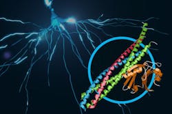
In this latest research, the scientists found that when the SNAREs and synaptotagmin-1 join up, they act as an amplifier for a slight increase in calcium concentration, triggering a gunshot-like release of neurotransmitters from one neuron to another. They also learned that the proteins join together before they arrive at a neuron’s membrane, which helps to explain how they trigger brain signaling so rapidly.
The team speculates that several of the joined protein complexes may group together and simultaneously interact with the same vesicle to efficiently trigger neurotransmitter release, an exciting area for further studies.
To study the joined protein structure, researchers in Brunger’s laboratory at the Stanford School of Medicine found a way to grow crystals of the complex. They used a robotic system developed at SSRL to study the crystals at SLAC’s LCLS, an x-ray laser that is one of the brightest sources of x-rays worldwide. The researchers combined and analyzed hundreds of x-ray images from about 150 protein crystals to reveal the atomic-scale details of the joined structure.
Brunger says that future studies will explore other protein interactions relevant to neurotransmitter release.
Full details of the work appear in the journal Nature; for more information, please visit http://dx.doi.org/10.1038/nature14975.
Follow us on Twitter, 'like' us on Facebook, connect with us on Google+, and join our group on LinkedIn