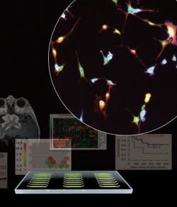
Microfluidics, cell imaging pair for single-cell molecular measurements
A microfluidic image cytometry (MIC) platform, developed at the University of California, Los Angeles (UCLA), makes in vitro measurements of intracellular signaling pathways and thus marks an advance in molecular diagnostics that could one day enable prognosis prediction and guide personalized treatment.1 "The MIC is essentially a cancer diagnostic chip that can generate single-cell 'molecular fingerprints' for a small quantity of pathology samples, including brain tumor tissues," said Hsian-Rong Tseng, associate professor of molecular and medical pharmacology and a leader of the research. "We are exploring the use of the MIC for generating informative molecular fingerprints from rare populations of oncology samples–for example, tumor stem cells. "The platform accommodates specimens with as few as 1,000 to 3,000 cells.
Led by Tseng and assistant professor Thomas Graeber, the 35-member interdisciplinary team analyzed a panel of 19 human brain tumor biopsies to demonstrate the platform's clinical application. "Because the measurements are at the single-cell level, computational algorithms are then used to organize and find patterns in the thousands of measurements," said Graeber, referencing the bioinformatics that the team devised. "These patterns relate to the growth signaling pathways active in the tumor that should be targeted in genetically informed or personalized anticancer therapies."
According to Nicholas Graham, a post-doctoral scholar at the California NanoSystems Institute (CNSI) who worked out the data analysis, "The single-cell nature of the MIC brain tumor data presented an exciting and challenging opportunity." To succeed, he said, their approaches needed to "preserve the power of single-cell analysis, but allow for comparison between patients."
The researchers will next apply the new platform to larger cohorts of cancer patient samples and integrate the diagnostic approach into clinical trials of molecular therapies. Recent graduates of UCLA's MBA program founded CytoScale Diagnostics in April to license the technology; the startup plans commercialization by the end of 2013.
1. J. Sun et al., Cancer Res 70: 6128-6138 (2010)
More BioOptics World Current Issue Articles
More BioOptics World Archives Issue Articles

