CELL BIOLOGY/LIVE CELL IMAGING: Laser-based quantitative measurement highlights role of cellular forces in developing tissue
Scientists at Harvard University's School of Engineering and Applied Sciences (SEAS) and Wyss Institute for Biological Engineering (Cambridge, MA) have used laser illumination to quantitatively measure in three dimensions (3D) the miniscule forces affecting developing cells in living embryos. The method could lead to new tools to diagnose cancer, hypertension, connective tissue diseases, and more: "Now that we can quantitate cellular forces, we can find entirely new ways to diagnose the extraordinarily wide range of diseases that alter cell contractility and tissue stiffness," says Don Ingber, founding director of the Wyss Institute, professor of bioengineering at SEAS, and senior author of the study. "Just as important, we can answer crucial questions about development that have lay dormant for decades."

Studies suggest that mechanical forces are as important in regulating biological function as chemicals and genes, but the study of such control mechanisms has been hampered by a lack of means to quantify mechanical forces at specific positions in living tissues. Such forces are particularly important as the body develops from the fertilized egg into tissues and organs with specialized shapes and functions—a process known as morphogenesis. Biologists studying morphogenesis knew that as the embryo develops, mechanical forces direct cells to multiply, steer to their proper locations, and specialize.
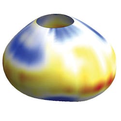
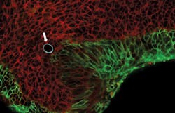
Otger Campás, assistant professor at the University of California, Santa Barbara (UCSB) and former postdoctoral fellow at SEAS and the Wyss Institute, had, as a doctoral student, used oil droplets to measure forces exerted by a network of protein filaments that drive cell movement. That work inspired his decision to use oil microdroplets as force transducers in living tissues. He, Ingber, and their colleagues identified a fluorocarbon that remains separate from the cell membrane, like oil does from water, and is safe for cells and tissues. Then, they devised a special coating for the droplets so it sticks to cells or to the extracellular matrix.
They coated the droplets with a chemical that made their surface glow when illuminated with a laser, then videotaped under a microscope as cells tugged and pressed on the droplet in 3D. By measuring how each droplet deformed, the scientists could precisely calculate the force exerted on it by the neighboring cells adhering to it and by the extracellular matrix.
Using lab-grown 3D aggregates of mouse mammary tumor cells and within living 3D tissues from embryonic mouse jaws, they found that an individual cell exerted huge forces—24 times as much pressure on the droplet as the jaws of an ant, and that the cells exerted the same amount of force in cultured aggregates as in tissues, lending confidence to the method's accuracy.
1. O. Campás et al., Nat. Meth., doi:10.1038/nmeth.2761 (2013).