MULTIPHOTON MICROSCOPY/CANCER DIAGNOSIS: Two-laser system improves melanoma diagnosis
A two-laser microscopy system developed at Duke University promises to help doctors more effectively diagnose melanoma. The technique claims to enable scientists to identify substantial chemical differences between cancerous and healthy skin tissues.
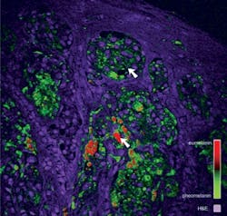
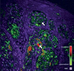
The two lasers pump small amounts of energy into suspicious moles; analysis of energy redistribution in the skin cells pinpoint the locations of different skin pigments. The approach enabled the Duke team to image 42 skin slices—which showed that melanomas tend to have more eumelanin, a kind of skin pigment, than healthy tissue. Using the amount of eumelanin as a diagnostic criterion, the team used the tool to correctly identify all 11 melanoma samples in the study.1
The technique will be further tested using thousands of archived skin slices. Studying old samples will verify whether the new technique can identify changes in moles that eventually did become cancerous. Current diagnostic approaches involve a light and magnifying glass or tissue biopsy—and are only about 85 percent accurate. So even if the technique proves, on a large scale, to be 50 percent more accurate than a biopsy, it would prevent about 100,000 false melanoma diagnoses, said Warren S. Warren, director of Duke’s Center for Molecular and Biomolecular Imaging, who oversaw the development of the new melanoma diagnostic tool.
The highly specialized lasers are currently commercially available and would only need to be added to the microscopes pathologists already use to diagnose melanomas. The cost for the added instrumentation is about $100,000, which may sound like a lot of money, but if each false-positive melanoma diagnosis costs thousands of dollars, having such an instrument available for questionable cases could considerably reduce health care costs overall, Warren said.
While the tool is designed for ex-vivo use, the researchers are investigating development of an in-vivo approach—which would not be ready for a few years.
1. T.E. Matthews, Sci. Transl. Med. 3 (71): 15 (2011).
More BioOptics World Current Issue Articles
More BioOptics World Archives Issue Articles

