LEDs enable fast, high-resolution multispectral eye imaging for disease screening
A new imaging system that uses six different wavelengths to illuminate the eye's interior (ocular fundus) may pave the way for doctors to easily screen patients for common diseases such as age-related macular degeneration (AMD) and diabetic retinopathy.1
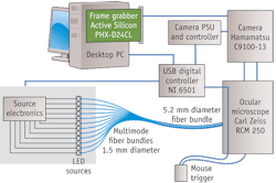
The current approach to eye imaging involves two or three wavelengths (red, green and blue) to highlight visually apparent abnormalities. The new system, developed by University College London researchers, allows doctors to distinguish between the different light-absorbing characteristics of chromophore molecules. Five light-absorbing compounds prevalent in the eye-retinal hemoglobins, choroidal hemoglobins, choroidal melanin, retinal pigment epithelium (RPE) melanin and macular pigment-give rise to distinct variations in tissue coloration that can be discriminated in multispectral images. The new device combines a high-sensitivity charge-coupled device (CCD) camera with wavelength-specific illumination from light-emitting diodes (LEDs) to enable multispectral imaging. The multispectral images are affected differently by the pigments present in the eye, and through a sophisticated algorithm they can be used to generate a pixel-by-pixel "parametric map" of the distribution of substances. Such maps may allow primary care clinicians to screen for and identify pathologies much earlier than before.
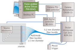
Unlike other multispectral retinal imaging devices, the new system can acquire a sequence of images fast enough (0.5 sec) to reduce image shifts caused by natural eye movements. And unlike the current snapshot systems, its images retain full spatial resolution. Because it uses only the specific wavebands required for the subsequent analysis, total light exposure is minimized-benefiting both patient safety and image quality.
1. N.L. Everdell et al., Rev. Sci. Instrum. 81: 093706 (2010)
More BioOptics World Current Issue Articles
More BioOptics World Archives Issue Articles