ADAPTIVE OPTICS/OPHTHALMOLOGY: First-ever clear, direct imaging of rod photoreceptors eyes ophthalmic advances

An innovation based on adaptive optics (AO) has made possible the first-ever clear and direct imaging of the tiny light-sensing cells in the living eye known as rods. Described in two journal papers in Optical Society (OSA) publications, the approach is expected to help ophthalmologists make earlier diagnosis of degenerative eye disorders, and lead to quicker intervention and more effective treatments.1,2
“It’s impossible to overemphasize how important early detection is to eye disease,” says researcher Alfredo Dubra of the University of Rochester in New York. (Dubra led the team of researchers from Rochester, Marquette University, and the Medical College of Wisconsin (MCW), Milwaukee.) “This is a really exciting breakthrough,” says Steve Burns, a professor in the School of Optometry at Indiana University, who is not involved in the work just published. “Imaging contiguous rod mosaics will allow us to study the impact of a whole new class of blinding disorders on the retina. Since many of the eye diseases most amenable to intervention affect the rods, this should become a major tool for determining what treatments work best for those disorders.”
“One of the major hurdles in detecting retinal disease is that by the time it can be perceived by the patient or detected with clinical tools, significant cellular damage has often already occurred,” adds team member Joseph Carroll of MCW.
Though earlier AO systems could effectively image cones and have become a mainstay of high-resolution retinal imaging research, the smaller rods, which outnumber cones 20 to 1 in the retina, have eluded clear and contiguous observation in the living eye.
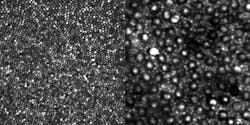
The design of the AO instrument that led to clearly visualizing rods was, according to Dubra, “embarrassingly simple, and relied on well-known equations and concepts.” By simply folding the spherical mirrors that act as lenses in the instrument into a three-dimensional structure, the image quality of the retina was improved sufficiently to clearly resolve the contiguous rod mosaic, as well as the entire cone mosaic at the foveal center. Dubra and his colleagues were able to push the device’s resolution to its optical limits of nearly 2 µm, or the approximate diameter of a single rod in the human eye.
According to the researchers, their next step is to develop a clinical model that could be widely available. A related task is simplifying and teaching the art of interpreting AO images to guide clinical decisions about diagnosis and treatment.
When that occurs, perhaps within the decade, doctors will likely be able to routinely peer into a living human eye with such precision and clarity that they will be able to see and evaluate individual rods—and do three things never before possible: accurately describe the physical presentation of specific rod disorders—the “phenotype” of a disease, intervene with early treatment at the first sign of disease, and even determine how individual cells are responding to a specific treatment.
1. A. Dubra et al., Biomed. Opt. Exp. 2 (7), 1864–1876 (2011).
2. A. Dubra and Y. Sulai, Biomed. Opt. Exp. 2 (6), 1757–1768 (2011).
More BioOptics World Current Issue Articles
More BioOptics World Archives Issue Articles