Optical probes overcome light-scattering issue in deep-brain imaging

Dec. 20, 2016
An implantable, ultra-narrow, silicon-based photonic probe can deliver light deep within brain tissues.

Light scattering and absorption in neural tissue cause light penetration to be extremely short, making it impossible to employ free-space optical methods like optogenetics to probe brain regions deeper than about 2 mm.

Related: Ten years after - Optogenetics progresses in clinical trials

Recognizing this, a team of researchers from the Calfornia Institute of Technology (Caltech; Pasadena, CA), the Baylor College of Medicine (Waco, TX), and Stanford University (CA) has developed a new approach that combines nanophotonics and microelectromechanical systems (MEMS) in an implantable, ultra-narrow, silicon-based photonic probe to deliver light deep within brain tissues. This minimally invasive technique avoids major tissue displacement during implantation.

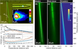
Characterization of E-pixel illumination, including an optical micrograph showing the side view of a shank immersed in a fluorescein–water solution to visualize the E-pixel illumination profiles (a), measured green PL intensity pattern, covering a distance of 410 μm, generated by the blue (473 nm) illumination beam emitted by the photonic E-pixel (b and c), simulated E-pixel illumination intensity profile in water (d), and PL beam profile analysis (e). (Image credit: E. Segev et al., Neurophoton., 4, 1, 011002 (Dec. 6, 2016), with permission from SPIE)

Using optogenetic techniques, a protein in the brain serves as a sensory photoreceptor and can be controlled by specific wavelengths of light. These combined techniques provide a new approach to stimulating brain circuits with remarkable resolution, enabling observation and control of individual neurons.

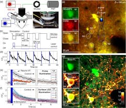
Cortical neural stimulation with concomitant two-photon optical functional imaging, including a schematic of the experimental setup (a), an illustration of the light excitation sequence (b), visualization of the expression levels of optogenetic actuators and reporters of the imaging site in mouse cortical layer (c), and the results from neural excitation (d-f). (Image credit: E. Segev et al., Neurophoton., 4, 1, 011002 (Dec. 6, 2016), with permission from SPIE)

These breakthroughs present widespread and promising applications for the neuroscience and neuromedical research communities. From characterizing the role of specific neurons and identifying neural circuits responsible for behavior to enabling new methods of operant conditioning through reward-induced circuit activations, optogenetics has become a new path for neuroscientists seeking advances in research capabilities.

Full details of the work appear in the journal Neurophotonics (open access); for more information, please visit http://dx.doi.org/10.1117/1.nph.4.1.011002.
Sponsored Recommendations

Hexapod 6-DOF Active Optical Alignment Micro-Robots - Enablers for Advanced Camera Manufacturing

Dec. 18, 2024
Optics and camera manufacturing benefits from the flexibility of 6-Axis hexapod active optical alignment robots and advanced motion control software

Laser Assisted Wafer Slicing with 3DOF Motion Stages

Dec. 18, 2024
Granite-based high-performance 3-DOF air bearing nanopositioning stages provide ultra-high accuracy and reliability in semiconductor & laser processing applications.

Steering Light: What is the Difference Between 2-Axis Galvo Scanners and Single Mirror 2-Axis Scanners

Dec. 18, 2024
Advantages and limitations of different 2-axis light steering methods: Piezo steering mirrors, voice-coil mirrors, galvos, gimbal mounts, and kinematic mounts.

Free Space Optical Communication

Dec. 18, 2024
Fast Steering Mirrors (FSM) provide fine steering precision to support the Future of Laser Based Communication with LEO Satellites

Voice your opinion!

To join the conversation, and become an exclusive member of Laser Focus World, create an account today!