Lab-scale laser device prototype discerns between healthy and damaged skin tissue
Researchers from the Technical University of Madrid, in collaboration with the Polytechnic University of Valencia and the Centro de Investigación Biomédica en Red (CIBER)'s Bioengineering, Biomaterials and Nanomedicine (CIBER-BBN; all in Spain), have designed a laser device specifically designed for optical hyperthermia applications, with the aim of easily distinguishing between healthy and damaged tissue.
The low-cost, lab-scale laser device was developed for treatments based on optical hyperthermia applications. This technique is used, for instance, in therapies against skin cancer, and it aims to kill the tumor cells by overheating them. Irradiating synthesized metallic nanoparticles heats the tumor tissue, reaching a temperature between 42° and 48°C to cause a hypoxia that leads to cellular death, explains Roberto Montes, a researcher from the Polytechnic University of Valencia.
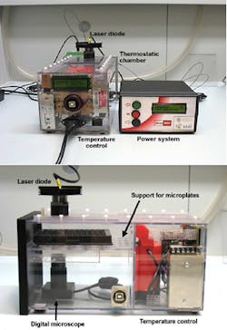
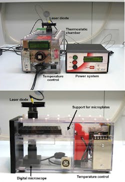
The prototype consists of an infrared (IR) laser with power up to 500 mW able to provide a power density up to 4 W/cm2, a sensor that records the temperature in real time during the irradiation, and a laser power regulator, among other components.
The device is already being used in in vitro cellular crops and in therapies that combine hyperthermia with the controlled release of drugs. "Although the equipment has been designed to exclusively work in a lab environment, once the technique is developed, it could be easily applied to a hospital environment with small changes," explains Javier Ibáñez from the Polytechnic University of Valencia.
Because the research team is in its initial phase with the device, Ibáñez says, they still need to conduct animal tissue testing, followed by testing on living animals and verifying its application in patients.
Full details of the work appear in the journal Sensors and Actuators A: Physical; for more information, please visit http://doi.org/10.1016/j.sna.2016.12.018.

